Understanding Ankle Injuries and Treatment Options
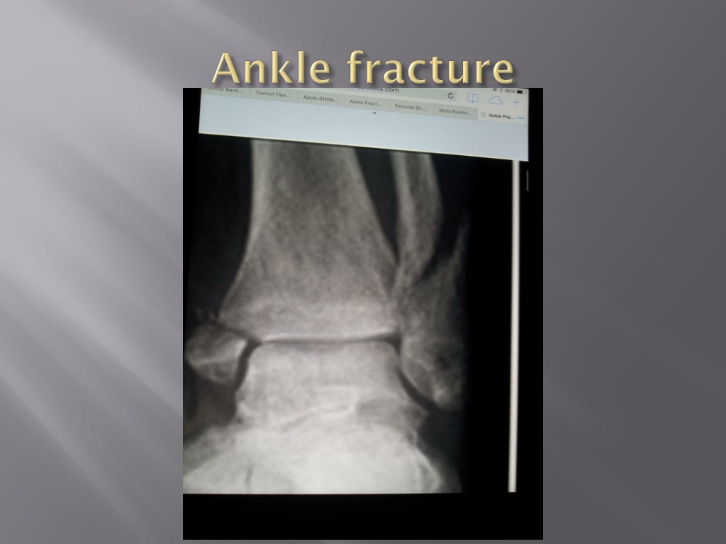
Explore the anatomy of deltoid ligaments, fibula, medial malleolus, and posterior malleolus in ankle injuries. Learn about deformities, swelling, and bruises associated with these injuries. Discover diagnostic techniques like radiographs and Lauge-Hansen classification. Delve into non-operative and operative management options, including short-leg casts, ORIF procedures, and plaster applications for fractures.

Download Presentation

Please find below an Image/Link to download the presentation.
The content on the website is provided AS IS for your information and personal use only. It may not be sold, licensed, or shared on other websites without obtaining consent from the author. Download presentation by click this link. If you encounter any issues during the download, it is possible that the publisher has removed the file from their server.
E N D
Presentation Transcript
Deltoid Ligaments Fibula Medial malleolus Posterior malleolus
1. Deformity 2. Swelling 3. Bruises 4. Antalgic gait
1. Radiographs External rotation stress radiograph Clear space >5mm - 2. AP view Lateral view Oblique view - -
Lauge-Hansen Supination adduction Supination external rotation Pronation aduction Pronation external rotation
A B C Lateral malleolar
Non-operative short-leg walking cast/boot <3mm displacement <25% pasterior malleolar fracture <2mm step-off - - - -
O.R.I.F Anatomic reduction -
Displaced O.R.I.F
3mm displaced O.R.I.F