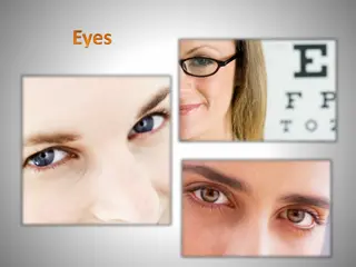
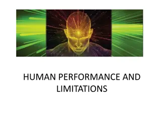
Exploring the Fascinating Physics of Eyes and Vision
Delve into the intricate workings of the human visual system, examining how our eyes perceive the world around us. Discover the remarkable features of our optical system, from automatic aperture adjustments to self-regulating pressure mechanisms. Uncover the three major components of vision and the crucial role each plays in shaping our perception. Gain insight into the exquisite design of the eye, which surpasses even the most advanced cameras in functionality. Explore the abilities of the eye, including its wide angle of observation, rapid focusing system, and dynamic muscle movements that enable flexible vision.

Download Presentation

Please find below an Image/Link to download the presentation.
The content on the website is provided AS IS for your information and personal use only. It may not be sold, licensed, or shared on other websites without obtaining consent from the author. Download presentation by click this link. If you encounter any issues during the download, it is possible that the publisher has removed the file from their server.
E N D
Presentation Transcript
Part One: - Physics of Eyes and Vision
Most of our knowledge of the world around us comes to us through our eyes. The helplessness we feel when caught in the dark in unfamiliar surroundings is a good indication of our dependence on vision.
The sense of vision consists of three major components: - 1. The eyes that focus an image from the outside world on the light-sensitive retina. 2. The system of millions of nerves that carries the information deep into the brain. 3. The visual cortex-that part of the brain where "it is all put together". Blindness results if any one of the parts does not function.
Our optical system has the following special features, most of which are not available on even the most expensive cameras: - 1. The eye can observe events over a very large angle while looking intently at an object directly ahead of it. 2. Blinking provides the front lens (cornea) with a built- in lens cleaner and lubricator. 3. A rapid automatic focusing system permits viewing objects as close as 20cm one second and distant objects the next. Under relaxed conditions the focus for normal eyes is set for "infinity" (distant viewing).
4. The eye can operate effectively over a range of light intensity of about 10 billion to one (1010:1)-brilliant daylight to very dark night. 5. The eye has an automatic aperture adjustment (the iris). 6. The cornea has a built-in scratch remover; even though it has no blood supply it is made of living cells and can repair local damage. 7. The eye has a self-regulating pressure system that maintains its internal pressure at about 20mmHg and thus keeps the eye in shape. If "dented", the eye rapidly returns to its original shape.
8. The eyes are mounted in a well-protected casing almost completely surrounded by bone, and each eye rests on a cushion of fat that reduces sharp shocks. 9. The image appears upside down on the light-sensitive retina at the back of the eyeball, but the brain automatically corrects for this. 10. The brain blends the images from both eyes, giving us good depth perception and true three-dimensional viewing. If vision from one eye is lost, the vision from the remaining eye is adequate for most needs. 11. The muscles of the eye permit flexible movement up and down, sideways, and diagonally. After a little practice, the eyes can even be made to go in circles.
Focusing elements of the eye The eye has two major focusing components: the cornea, which is the clear transparent bump on the front of the eye that does about two-thirds of the focusing, and the lens, which does the fine focusing. The cornea is a fixed focus element; the lens is variable in shape and has the ability to focus objects at various distances. The cornea focuses by bending (refracting) the light rays. The amount of bending depends on the curvatures of its surfaces and the speed of light in the lens compared with that in the surrounding material (relative index of refraction).
When the cornea is under-water it loses most of its focusing power because the index of refraction of the water (1.33) is close to that of the cornea (1.37). Fish have a similar problem out of the water. Divers keep air around the cornea by wearing a face mask. Nearly all of the focusing by the cornea is done at the front surface since the aqueous humor in contact with the back surface has nearly the same index of refraction as the cornea. Since the living cells in the cornea are not supplied with oxygen by the blood, they must get their oxygen from the air. Having blood vessels in the cornea would not help our vision! The nutrients for the cells in the cornea are supplied by the aqueous humor that is in contact with its back surface. The aqueous humor contains all of the blood components except blood cells.
If the cornea is scratched it will heal itself, but some other types of damage are more permanent. Some types of radiation (ultraviolet, neutrons, X-rays, etc.) can cause opacities to develop in the cornea that will block out light. The lens has focusing properties at both its front surface and its back surface. The lens is more curved in the back than in the front. It changes its focal strength by changing its curvature. The focusing power of the lens is considerably less than that of the cornea because it is surrounded by substances that have indexes of refraction close to its own. The lens is made up of layers somewhat like an onion, and all layers do not have the same index of refraction. The lens, like the cornea, can be damaged by ultraviolet and other forms of radiation. It can develop cataracts, which destroy its clarity.
Some other elements of the eye The pupil is the opening in the center of the iris where light enters the lens. It appears black because essentially all of the light that enters is absorbed inside the eye. Under average light conditions, the opening is about 4mm in diameter. It can change from about 3mm in diameter in bright light to about 8mm in diameter in dim light. It is believed that the iris aids the eye by increasing or decreasing incident light on the retina until the retina has adapted to the new lighting conditions. In addition, under bright light conditions it plays an important role in reducing lens defects.
The aqueous humor fills the space between the lens and the cornea. This fluid, mostly water, is continuously being produced, and the surplus escapes through a drain tube, the canal of Schlemm. Blockage of the drain tube results in increased pressure in the eye; this condition is called glaucoma. The aqueous humor contains many of the components of blood and provides nutrients to the nonvascularized cornea and lens. It maintains the internal pressure of the eye at about 20mmHg. If you press on the eye, you find it is fairly stiff; you cannot indent it much. The reasons are that the fluids in the eye are incompressible at the pressure you use and that the covering of the eyeball does not stretch easily. When you rub your eyes, you greatly increase the internal pressure.
The vitreous humor is a clear jelly-like substance that fills the large space between the lens and the retina. It helps keep the shape of the eye fixed and is essentially permanent. It is sometimes called the vitreous body. The sclera is the tough, white, light-tight covering over all of the eye except the cornea. The sclera is protected by a transparent coating called the conjunctiva.
The retina-the light detector of the eye The retina, the light-sensitive part of the eye, converts the light images into electrical nerve impulses that are sent to the brain. The absorption of a light photon in a photoreceptor triggers an electrical signal to the brain-an action potential. The energy of the photon is about 3eV; the action potential has an energy millions of times greater. The light photon apparently causes a photochemical reaction in the photoreceptor which in some way initiates the action potential. The photon must be above a minimum energy to cause the reaction. Infrared photons have insufficient energy and thus are not seen. Ultraviolet photons have sufficient energy, but they are absorbed before they reach the retina and also are not seen.
The retina covers the back half of the eyeball. While this large expanse permits useful "warning" vision over a large angle, most vision is restricted to a small area called the macula lutea, or yellow spot. All detailed vision takes place in a very small area in the yellow spot (~0.3mm in diameter) called the fovea centralis. The image on the retina is very small. A convenient equation for determining the size of the image on the retina comes from the ratios of the lengths of the sides of similar triangles. O is the object size, I the image size, P the object distance, and Q the image distance.
* How big is the image on the retina of a fly on a wall 3.0 m away? Assume the fly is 3 mm (0.003 m) in diameter and Q = 0.02 m. O P = I Q Q = I xO P . 0 02 = = = 5 . 0 003 2 10 20 I x x m m 3
There are two general types of photoreceptors in the retina: the conesand the rods. Throughout most of the retina the cones and rods are not at the surface of the retina but lie behind several layers of nerve tissue through which the light must pass. However, in the fovea centralis most of this nerve tissue is pushed to the side and there is a slight dip. The rods and cones are distributed symmetrically in all directions from the visual axis except in one region-the blind spot.
The cones (~6.5 million in each eye) are primarily used for daylight, or photopic,vision. With the cones, we can see fine detail and recognize different colors. The cones are primarily found in the fovea centralis, although some are scattered throughout the retina. We see later that the density of the cones in the fovea centralis determines the amount of detail we can resolve in an image. Each of the cones in the fovea has its own "telephone line" to the brain. In the rest of the retina several cones share one nerve fiber. The cones are not uniformly sensitive to all colors but have a maximum sensitivity at about 550nm in the yellow-green region.
The rods are used for night, or scotopic, vision and for peripheral vision. They are much more abundant than the cones (~120 million in each eye) and cover most of the retina. They are not uniformly distributed over the retina but have a maximum density at an angle of about 20o. Histological studies have indicated that hundreds of rods send their information to the same nerve fiber. The rods are most sensitive to blue-green light (~510nm), which has a wavelength shorter than the optimum for the cones (~550nm). The rods and cones are equally sensitive to red light (650 to 700 nm).
The eyes do not have their greatest sensitivity to light under photopic conditions. If the light level suddenly decreases by a factor of 1000 we are momentarily "in the dark", but after a few minutes we are able to see many of the details that were not visible when it first became dark. This dark adaptation is apparently the time needed for the body to increase the supply of photosensitive chemicals to the rods and cones. The cones adapt most rapidly; after about 5min the fovea centralis has reached its best sensitivity. The rods continue to dark adapt for 30 to 60 min, although most of their adaptation occurs in the first 15min. There is a region from about 13o to 18o that has neither rods nor cones-the blind spot. This is the point at which the optic nerve enters the eye. The blind spot is on the side toward the nose.
How little light can you see? If 90 photons enter the eye, 10 or less are actually absorbed in photoreceptors. What happens to the others? About 3% are reflected at the surface of the cornea, and about 50% are absorbed in the various structures (cornea, lens, humors). Of those that reach the region of the rods, only about 20% (~10% of the original number) are absorbed in the rods. The photons that miss the rods are absorbed in the "backstop". Some animals, such as cats, have a reflective coating behind the rods that gives the rods another opportunity to absorb the photons. These animals have eyes that "glow in the dark" if a light shines in them.
Diffraction effects on the eye All light waves undergo diffraction as they pass through a small opening. Thus the iris produces a diffraction pattern on the retina. At the normal opening of the pupil (~4mm) this phenomenon has no practical consequences for our daily vision tasks. However, if the pupil becomes much smaller, for example, 1.0mm, diffraction produces a measurable effect on visual acuity. All lenses have defects (aberrations). The effect of such aberrations is reduced if the lens opening is made smaller. In the eye, a small pupil improves visual acuity. However, if the pupil is made very small the acuity becomes worse due to diffraction effects.
How sharp are your eyes? The familiar eye charts used to determine whether we need corrective lenses test the property of our eyes called visual acuity. A physicist calls visual acuity the resolution of the eyes. The optometrist usually uses a Snellen Chart to test visual acuity. If he tells you that your eyes test normal at 20/20, he means that you can read detail from 20ft that a person with good vision can read from 20ft. If your eyes test at 20/40, you can just read from 20ft the line that a person with good vision can read from 40ft. The Snellen Chart tests many things besides acuity.
The visual acuity or resolution of the eye is primarily determined by the characteristics of the cones in the fovea. A common way of testing resolution is to use a pattern of alternating black and white lines that become increasingly narrower. A combination of one white line and one black line is called a line pair (lp). Under optimum conditions, the eye can just barely resolve as separate lines a pattern of about 30 lp/mm; when the eye is twice as far away, it can only resolve 15 lp/mm. The resolution rapidly gets worse as the image moves away from the fovea centralis. If the lighting is not optimum the resolution also deteriorates.
One test of acuity used often by scientists is aligning two lines so that they appear to be a single continuous line. This is involved in using a slide rule and also in using a vernier scale, and it is referred to as vernier acuity. Light letters on a dark background are not as easy to read as the conventional dark letters on a light background. The ability of the eye to recognize separate lines also depends on the relative "blackness" and "whiteness" of the lines. Resolution is much worse if the lines are two close shades of gray than if one is black and one is white. The contrast (C) between two areas is defined as:-
I ( I ) = C 1 2 + I ( I ) 1 2 Where I1 and I2are the light intensities from the two areas. The low contrast between two areas of interest on an X-ray often severely limits the usefulness of the X-ray. Before discussing contrast further, we need to define a unit for measuring the "darkness" of an X-ray film or any other optical absorber. The optical density ODis defined as: - log( OD = I ) o I Io is the light intensity without the absorber. I is the intensity with the absorber.
* A piece of film that transmits 10% of the incident light has an OD? 100 I o = = = 0 . 1 = log( ) log( ) log 10 OD 10 I * What is the amount of light transmitted by a film of OD=0.5? I I = = o o OD log( ) or antilog OD I I I = = o antilog 0.5 3.15 I I = o I , or about one third of the light is transmitte d. 3.15 The range of optical densities used for most X-ray viewing is (0.3-2.0).
Optical illusions and related phenomena Viewing shades of gray plays tricks on the mind. A given bar looks darker where it borders a lighter bar and lighter on the side near a darker bar. This effect is produced by interactions between adjacent groups of light sensors in the eye. This "edge enhancement" would appear to help radiologists see borders on X-ray films. Another optical illusion is that the shade an area of gray appears depends on its surroundings. The circle surrounded by a light area looks darker. This effect has practical significance for viewing X-ray films on an illuminator.
If you look into the eye with an ophthalmoscope you can see the many blood vessels in the retina. These blood vessels block out light to the rods and cones behind them. The reason we do not normally see these vessels is that the shadows they produce are always over the same rods and cones and this steady signal fades in moments after we open our eyes in the morning. Everyone has at one time or another seen "lights" when his eyes were closed. These "lights" are called phosphenes and can be stimulated by pressing on the eye with the fingers or by closing the eyes very tightly. Phosphenes are produced by the stimulation of some of the normal light sensors. The brain interprets any signals received from the optic nerve as light; it cannot distinguish the various sources of signals from the optic nerve.
A characteristic of the eye-brain system that we take for granted is the ability of the brain to fuse the slightly different images from the eyes into a three-dimensional (3-D) image. An artificial 3-D effect can be produced by showing slightly different pictures to the two eyes. About 150 years ago Wheatstone invented the stereoscope, which is still the basic device used to view stereoscopic X-rays. For stereoscopic X-rays, two X-rays are taken of the same part of the body from slightly different angles corresponding to the views normally seen by the two eyes. The two X-rays are then placed in a stereoscope so that each eye sees one image; these images are fused by the brain into a 3-D image. Stereoscopic X-rays are often taken of the skull.
It is also possible to take 3-D pictures in an electron microscope by tilting the specimen slightly before taking the second picture. This technique can be used to see inside a chromosome. When you view a flash of light, the visual image of the light persists for some time after the flash. That is, after the flash there is a period of many milliseconds when the brain thinks the light is still on.


 are primarily")