Understanding Temperature Measurement in Healthcare

Temperature, Pulse, and Respirations are vital signs used to assess a patient's health. Temperature can be measured through various methods such as oral, rectal, axillary, tympanic, and temporal routes using different types of thermometers. It's important to know the normal temperature ranges for different measurement sites and understand temperature terms like hypothermia, fever, and hyperthermia. Certain precautions need to be taken when measuring temperatures in specific patient populations.

Download Presentation

Please find below an Image/Link to download the presentation.
The content on the website is provided AS IS for your information and personal use only. It may not be sold, licensed, or shared on other websites without obtaining consent from the author. Download presentation by click this link. If you encounter any issues during the download, it is possible that the publisher has removed the file from their server.
E N D
Presentation Transcript
TPR Temperature, Pulse and Respirations
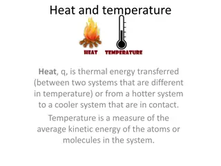
Temperature Is the measurement of the balance between heat lost and heat produced by the body
Temperature Can be measured by four basic routes 1. Oral Mouth- leave in place for 3-5 minutes 2. Rectal Rectum- leave in place for 3-5 minutes 3. Axillary Axilla or groin- leave in place for 10 minutes 4. Tympanic Eardrum- 5. Temporal Across forehead-
Types of Thermometers 1. Electronic/Digital 2. Glass 3. Thermoscan for Tympanic measurement 4.Temporal measurement thermometers
Normal temperature ranges Oral 97.6 F 99.6 F (36.5-37.5 C) Axillary or Groin 96.6 F 98.6 F ( 36- 37 C) one degree Fahrenheit lower than Oral Rectal & Temporal 98.6 F 100.6 F (37-38.1 C) one degree Fahrenheit higher than Oral
Normal Temperature Ranges Rectal & Temporal 98.6 F 100.6 F (37-38.1 C) one degree Fahrenheit higher than Oral Aural or Tympanic An ear (tympanic) temperature is 0.5 F (0.3 C) to 1 F (0.6 C) higher than an oral temperature--- 98.1- 100.1 F ( 36.8- 37.8 C)
Need to Know-Temperature Terms Hypothermia Below 95F ( 35C) Death at 93F (33.9) Fever Elevated above 101 (38.3) Pyrexia= febrile= fever present Afebrile= normal temp or no fever present Hyperthermia Temp exceeds 104 F (40C) Convulsions & death at 106 F ( 41.1 C)
Do not take oral temperatures on preschool children patients with oxygen delirious, confused, disoriented patients comatose patients patients with nasogastric tubes in place patients who have had oral surgery patients who are vomiting or nauseated
Do not take rectal temperatures on infants or children unless a core temperature is needed patients who have had rectal surgery combative patients
Abnormal temperatures Fever, febrile, hyperthermia all indicate someone who has an elevated temperature (>100 Fahrenheit). High fever would include anything over 103 degrees Fahrenheit. Moderate fever would include anything 100 103 degrees Fahrenheit. Hypothermia (<96F)is subnormal temperature. This can be equally problematic for a person
Need to Know Conversion Formulas Fahrenheit to Celsius C=(F-32)/ 1.8 Celsius to Fahrenheit F=(C X 1.8) + 32
Pulse **Student will learn how to asses pulses **
Assessing Temperatures With a partner Take both an oral and axillary temperature using a digital thermometer Record each temperature reading in both Fahrenheit and Celsius using the correct formula Take a tympanic temperature Document your temperature
Pulse Wave of blood produced and felt along the artery when the heart contracts and rests ( relaxes) BEATS Can feel at points where the artery is between finger tips and a bony area
Need to Know Pulse Terms Rate Number of bests/per minute Rhythm Regularity of the pulse Volume Refers to the strength of the pulse Apical pulse Pulse take at the apex of the heart with a stethoscope
Pulse Points- NEED TO KNOW 1. Temporal --either side of forehead 2. Carotid- at neck- either side of trachea 3. Apical- at apex of heart 4. Brachial-inner aspect of antecubital space 5. Radial- inner aspect of the wrist 6. Femoral- inner aspect of the upper thigh where it meets trunk-- groin 7. Popliteal- behind the knee 8. Dorsal Pedis -at the top of the foot arch
Measuring Pulses Measured by index, middle, and ring fingers over pulse point. Do not take with the thumb, since it has a pulse of its own. Count for 30 seconds and multiply by 2, or count for 60 seconds
Pulse Ranges Normal = Adults ----- 60 -100 beats/minute Children 7 year & older --- 65-80 /minute Children 1- 7 years--------- 80-110/ minute Infants birth 1 year-------100-160/minute > than 100 = tachycardia < than 60 = bradycardia
Quality of Pulse Rhythm regular or irregular Strength Bounding or thready
What do you think???? Jot down at least 5 factors that you think may contribute to your pulse rate accelerating decelerating
Circumstances affecting pulse rate 1. Body temperature 2. Emotions 3. Activity level 4. Health of heart 5. Medication 6. Sleep 7. Coma 8. Exercise 9. Shock states
Assessing Pulses Pick a partner Assess the following pulses for one full minute Record rate, rhythm, volume of the pulse Temporal Carotid Apical Brachial Radial Popliteal Dorsalis pedis Repeat all pulses after your partner has done 25 jumping jacks
Respirations Process of taking in O2 and expelling CO2 one respiration consists of One inspiration One expiration Please note the following when mearusing each and every respiration: 1. Rate 2. Character 3. Rhythm
Respirations Each breath includes inspiration and expiration. Measure by observing chest rise and fall. Measured in breaths per minute.
Respirations Rate number of breaths/ minute Character Depth and quality of respirations Deep-shallow-difficult-stertorous-moist Rhythm Regularity of respirations
Need to Know Respiration Terms Dyspnea Difficult or labored breathing Apnea Absence of respirations Tachypnea Rapid, shallow respirations-- < 25/minute Bradypnea Slow respiratory rate- > 10/minute Orthopnea Difficulty breathing in all positions except sitting or standing
Need to Know Terms Cheyne- stokes Abnormal respirations in a dyspnea and apnea pattern Rales Noisy & bubbling Wheezing Difficult breathing with high pitch whistling Cyanosis Dusky, bluish discoloration of skin, lips, nail beds
Ranges in Respirations Normal = adults12-24 breaths per minute Children-16-30/ minute Infants- 30-50/ minute > than 24 = tachypnea if breathing in great depth then called hyperpnea < than 12 = bradypnea Assess rate, character and rhythm always!!!
Quality of breathing 1. Depth 2. Clarity of breath sounds 3. Pain with breathing 4. Difficulty breathing use of accessory muscles
Assessing Respirations Assess the radial pulse rate of the patient for one minute After the pulse rate have been counted leave your hand in the pulse position Count the number of respirations- chest rise and fall for one minute Each complete cycle is ONE respiration
Pulse Oximetry Pulse oximetry is a procedure used to measure the oxygen level (or oxygen saturation) in the blood. It is considered to be a noninvasive, painless, general indicator of oxygen delivery to the peripheral tissues (such as the finger, earlobe, or nose).
How it works. Pulse oximetry technology uses the light absorptive characteristics of hemoglobin & the pulsating nature of blood flow in the arteries to aid in determining the oxygenation status in the body There is a color difference between arterial hemoglobin saturated with oxygen, which is bright red, and venous hemoglobin without oxygen, which is darker. with each heartbeat there is a slight increase in the volume of blood flowing through the arteries Pulse Oximetry measures the maximum amount of oxygen-rich hemoglobin pulsating through the blood vessels
Normal / Abnormal Values Normal pulse oximeter readings range from 95 to 100 percent, under most circumstances Values under 90 percent are considered low Hypoxemia describes a lower than normal level of oxygen in your blood.
Pain Assessment Pain is subjective Pain is also multidimensional, so the clinician must consider multiple aspects (sensory, affective, cognitive) of the pain experience. the nature of the assessment varies with multiple factors so no single approach is appropriate for all patients or settings.
Pain Assessment Onset & duration Location Quality-what does it feel like? Intensity- give a numeric reading Alleviating or exacerbating factors
Common Assessment Tools Wong Baker Scale Numeric Scales