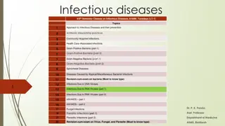
Viral Specimen Selection and Collection Overview
Specimen selection for viral testing depends on the suspected viral etiologies and disease syndrome. Throat and nasopharyngeal swabs are used for different viruses, while rectal swabs, urine, blood, and tissue samples are also collected for specific viral detection. Proper specimen transport and storage are crucial for accurate testing results, including using viral transport media and maintaining appropriate temperatures. Diagnostic methods include direct detection, virus isolation, and serology to detect viral particles, antigens, and nucleic acids.
Download Presentation

Please find below an Image/Link to download the presentation.
The content on the website is provided AS IS for your information and personal use only. It may not be sold, licensed, or shared on other websites without obtaining consent from the author.If you encounter any issues during the download, it is possible that the publisher has removed the file from their server.
You are allowed to download the files provided on this website for personal or commercial use, subject to the condition that they are used lawfully. All files are the property of their respective owners.
The content on the website is provided AS IS for your information and personal use only. It may not be sold, licensed, or shared on other websites without obtaining consent from the author.
E N D
Presentation Transcript
Viral Specimen Selection and Collection: Specimen selection depends on the specific disease syndrome, viral etiologies suspected and time of year. 1.Throat, nasopharyngeal swab or aspirate: Throat swabs are acceptable for recovering enteroviruses, adenoviruses and HSV, whereas nasopharyngeal swab or aspirate specimens are preferred for the detection of influenza and parainfluenza viruses. Throat specimens are collected by rubbing inflamed, vesiculated, or purulent areas of the posterior pharynx with a dry, sterile swab. Nasopharyngeal secretion specimens are collected by inserting a swab with flexible shaft through the nostril to the nasopharynx.
2.Rectal swabs and stool specimens: Used to detect rotavirus, enteric adenoviruses and enteroviruses. Rectal swabs are collected by inserting a swab (3-5) cm into the rectum to obtain feces. 3.Urine: CMV, mumps, rubella, measles, polyomaviruses and adenoviruses can be detected in urine. Virus recovery may be increased by processing multiple (2-3) specimens because virus can be shed intermittently or in low numbers. The best specimen is at least (10 ml) of a clean-voided first-morning urine. 4.Blood: Used to detect CMV, HSV, enterovirus and adenoviruses. (50-10 ml) of anticoagulated blood collected in a vacutaner tube is needed. 5.Tissue: Useful for detecting viruses that infect lung (CMV, influenza virus, adenovirus), brain (HSV), and gastrointestinal tract (CMV). Specimens are collected during surgical procedures.
Specimen transport and storage: 1-All specimens collected for detection of viruses should be collected with aseptic technique and transported immediately to the laboratory by using transport media (in screw capped tube) and should be placed in ice, specimens for viral isolation should not be kept at room or higher temperature. Under unusual circumstances, specimens may need to be held for days before processing. For storage up to 5 days, hold specimen at 4 C. Storage for 6 or more days should be at 20 C or preferably at 70 C.
2-Some antibiotics (e.g penicillin, streptomycin, nystatin) should be added to viral transport media especially when contamination with microbial flora is expected. Examples of successful transport media include Stuart s medium, Amie s medium. 3-Centrifuge under refrigeration, the supernatant will contain the viruses. 4-Filter the supernatant to give crude liquid of viruses. 5-Detection of viruses in this crude liquid of viruses by 3 methods
Overview of diagnostic methods In general, diagnostic tests can be grouped into 3 categories.: (1) direct detection, (2) indirect examination (virus isolation), and (3) serology. In direct examination, the clinical specimen is examined directly for the presence of virus particles, virus antigen or viral nucleic acids. In indirect examination, the specimen into cell culture, eggs or animals in an attempt to grow the virus: this is called virus isolation. Serology actually constitute by far the bulk of the work of any virology laboratory. A serological diagnosis can be made by the detection of rising titres of antibody between acute and convalescent stages of infection, or the detection of IgM. In general, the majority of common viral infections can be diagnosed by serology. The specimen used for direction detection and virus isolation is very important. A positive result from the site of disease would be of much greater diagnostic significance than those from other sites. For example, in the case of herpes simplex encephalitis, a positive result from the CSF or the brain would be much greater significance than a positive result from an oral ulcer, since reactivation of oral herpes is common during times of stress.
1. Direct Examination of Specimen Electron Microscopy morphology / immune electron microscopy Light microscopy histological appearance - e.g. inclusion bodies Antigen detection immunofluorescence, ELISA etc. Molecular techniques for the direct detection of viral genomes 2. Indirect Examination Cell Culture - cytopathic effect, haemadsorption, confirmation by neutralization, interference, immunofluorescence etc. Eggs pocks on CAM - haemagglutination, inclusion bodies Animals disease or death confirmation by neutralization
3. Serology Detection of rising titres of antibody between acute and convalescent stages of infection, or the detection of IgM in primary infection. Classical Techniques 1. Complement fixation tests (CFT) 2. Haemagglutination inhibition tests 3. Immunofluorescence techniques (IF) 4. Neutralization tests 5. Single Radial Haemolysis Newer Techniques 1. Radioimmunoassay (RIA) 2. Enzyme linked immunosorbent assay (EIA) 3. Particle agglutination 4. Western Blot (WB) 5. Recombinant (RIBA), line immunoassay (Liatek) etc. immunoblot assay


