Three-Dimensional Trochlear Groove Curvilinearity vs. Tibial Tubercle-Trochlear Groove Distance in Predicting Patellofemoral Instability
Patellofemoral joint stability relies on various factors, with the TT-TG distance commonly used to assess patellar instability risk. However, a study suggests that 3D measurements of trochlear groove curvilinearity may be more effective in differentiating individuals with recurrent patella dislocation from controls. The research compared these metrics in a cohort of patients with recurrent patellar dislocation and control subjects, using CT scans and 3D models. Findings indicate potential clinical implications for assessing patellofemoral instability risk.

Download Presentation
Please find below an Image/Link to download the presentation.
The content on the website is provided AS IS for your information and personal use only. It may not be sold, licensed, or shared on other websites without obtaining consent from the author. Download presentation by click this link. If you encounter any issues during the download, it is possible that the publisher has removed the file from their server.
Presentation Transcript
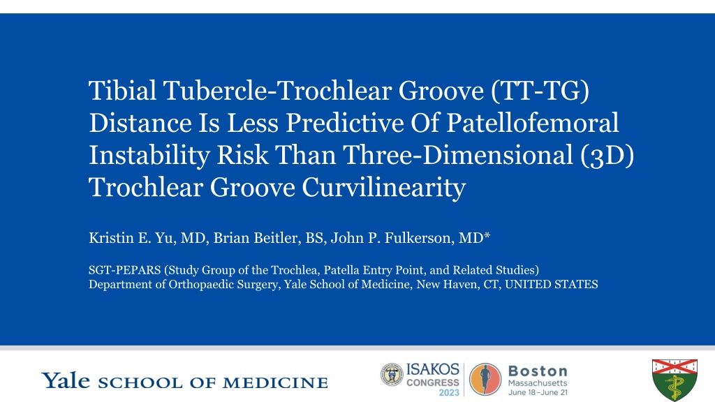
Tibial Tubercle-Trochlear Groove (TT-TG) Distance Is Less Predictive Of Patellofemoral Instability Risk Than Three-Dimensional (3D) Trochlear Groove Curvilinearity Kristin E. Yu, MD, Brian Beitler, BS, John P. Fulkerson, MD* SGT-PEPARS (Study Group of the Trochlea, Patella Entry Point, and Related Studies) Department of Orthopaedic Surgery, Yale School of Medicine, New Haven, CT, UNITED STATES S L I D E 1
Disclosures Kristin E. Yu, MD: none Brian Beitler, BS: none John P. Fulkerson, MD Paid Consultant for Conmed Support received from DJ Ortho Royalties or support received from Walters Kluwer Board of Directors member for Patellofemoral Foundation S L I D E 2
Background Patellofemoral joint (PFJ) stability dependent on static and dynamic stabilizers(1-7) Tibial tubercle-trochlear groove (TT-TG) distance commonly used in evaluation of patellar instability High inter- and intra-rater reliability(5, 8-9) Predictive of risk of future patellar dislocation and instability events(10-12) Range of normal values, surgical cutoffs debated(13-21) TT-TG distance affected by age, height, presence of trochlear dysplasia(13, 18-21) Isometry of medial patellofemoral ligament (MPFL) reconstruction changes at lower TT-TG distances in presence of patella alta(16, 22-23) Relationship between coronal malalignment and dysplasia remains unclear(16, 24) Objective: To compare the TT-TG distance in differentiating between subjects with recurrent patella dislocation and controls in comparison to a three-dimensional (3D) measurement of trochlear groove curvilinearity, the entry point (EP)-transition point (TP) angle. S L I D E 3
Methods N=24 (18F, 6M) patients with recurrent patellar dislocation (RPD) from Jan 2020-Nov 2021 Exclusion criteria: congenital deformity, history of knee trauma, arthritic disease CT scans of knee obtained in full extension and 30 degrees of flexion Whole body CTs obtained from n=10 control subjects, all female Sourced from New Mexico Decedent Image Database (NMDID) No history of patellar instability events or knee trauma Bilateral knees segmented and printed (n=20 control knees) 3D models printed 100% to scale Segmentation via Simpleware ScanIP (Synopsys, Mountain View, CA) Form 3BL printer (Formlabs, Somerville, MA), 0.1 mm resolution Trochlear groove curvilinearity compared between control and patellar dislocation cohorts TT-TG distances and trochlear widths (TW) measured on axial CT cuts Entry point (EP)-transition point (TP) angle(24) measured on 3D prints Measurements obtained with models resting on posterior femoral condyles Statistical analyses in Prism 9.0 S L I D E 4
Methods Entry point (EP): midpoint of flattened region of proximal trochlea that first engages with patella in early knee flexion Transition point (TP): point along trochlear groove at which direction of patellar tracking changes from oblique orientation proximally to vertical towards intercondylar notch distally EP-TP angle as shown in an age-matched control knee model (left) and a recurrent patellar dislocation knee model. An EP-TP angle of 141.3 was measured in the control knee, as compared with an EP-TP angle of 120.1 in the dislocator knee. Example EP-TP angles in six patients with recurrent patellar dislocations, shown on virtual 3D models. TPs are demarcated with a yellow star on each model. Figure from Yu, Kristin, Cooperman, DR, Schneble, CA, McLaughlin, W, Beitler, B, Kaliney, R, Wang, A, Fulkerson, JP. Reconceptualization of Trochlear Dysplasia in Patients With Recurrent Patellar Dislocation Using 3-Dimensional Models. Orthopaedic Journal of Sports Medicine. 2022; 10(11):232596712211382. S L I D E 5
Results Average age of RPD cohort: 20.3 yrs (range: 15-43) Average age of control cohort: 19.7 yrs (range: 15-25) No significant difference in TT-TG distance between cohorts by two-way, unpaired student s t-test (p=0.1424) No significant difference with normalization for trochlear width (TW), p=0.2491 TT-TG cutoff >14.9 mm on CT LR+ 2.333 for recurrent instability Sensitivity 77.8% Specificity 66.7% S L I D E 6
Results Significant difference in EP-TP angles between control and RPD cohorts (p<0.0001) Larger, more obtuse angles in control subjects Highest LR+ for recurrent instability at EP-TP angle cutoff <145 EP-TP angles <145 16x more likely in RPD cohort Sensitivity 80% Specificity 95% S L I D E 7
Discussion EP-TP angle demonstrates superior discriminative capacity between patients with RPD and controls over the TT-TG distance TT-TG accuracy for patellar instability increasingly debated, especially with trochlea dysplasia EP-TP good inter- and intra-rater reliability(24) Study limitations Sample size Comparison of disparate visualization modalities (2D CT scans vs 3D prints) Additional work needed to validate EP-TP angle cutoff S L I D E 8
References 1. 2. 3. 4. 5. 6. 7. 8. 9. Dejour H, Walch G, Nove-Josserand L, Guier C. Factors of patellar instability: an anatomic radiographic study. Knee Surg Sports Traumatol Arthrosc. 1994;2:19-26. Amis AA. Current concepts on anatomy and biomechanics of patellar stability. Sports Med Arthrosc Rev. 2007;15:48-56. Arendt EA, Fithian DC, Cohen E. Current concepts of lateral patella dislocation. Clin Sports Med. 2002;21:499-519. Bollier M, Fulkerson JP. The role of trochlear dysplasia in patellofemoral instability. J Am Acad Orthop Surg. 2011;19:8-16. Levy BJ, Tanaka MJ, Fulkerson JP. Current Concepts Regarding Patellofemoral Trochlear Dysplasia. Am J Sports Med. 2020:363546520958423. Sherman SL, Plackis AC, Nuelle CW. Patellofemoral anatomy and biomechanics. Clin Sports Med. 2014;33:389-401. Senavongse W, Amis AA. The effects of articular, retinacular, or muscular deficiencies on patellofemoral joint stability: a biomechanical study in vitro. J Bone Joint Surg Br. 2005;87:577-582. Pace JL, Cheng C, Joseph SM, Solomito MJ. Effect of Trochlear Dysplasia on Commonly Used Radiographic Parameters to Assess Patellar Instability. Orthop J Sports Med. 2020;8:2325967120938760. Stepanovich M, Bomar JD, Pennock AT. Are the Current Classifications and Radiographic Measurements for Trochlear Dysplasia Appropriate in the Skeletally Immature Patient? Orthop J Sports Med. 2016;4:2325967116669490. Tanaka MJ, D'Amore T, Elias JJ, Thawait G, Demehri S, Cosgarea AJ. Anteroposterior distance between the tibial tuberosity and trochlear groove in patients with patellar instability. Knee. 2019;26:1278- 1285. Clark D, Metcalfe A, Wogan C, Mandalia V, Eldridge J. Adolescent patellar instability: current concepts review. Bone Joint J. 2017;99-B:159-170. Laidlaw MS, Diduch DR. Current Concepts in the Management of Patellar Instability. Indian J Orthop. 2017;51:493-504. Camp CL, Heidenreich MJ, Dahm DL, Stuart MJ, Levy BA, Krych AJ. Individualizing the Tibial Tubercle-Trochlear Groove Distance: Patellar Instability Ratios That Predict Recurrent Instability. Am J Sports Med. 2016;44:393-399. Moya-Angeler J, Vairo GL, Bader DA, Sebastianelli WJ, Sherbondy PS. The Tibial Tubercle-Trochlear Groove Distance/Trochlear Dysplasia Index Quotient Is the Most Accurate Indicator for Determining Patellofemoral Instability Risk. Arthroscopy. 2022;38:1608-1614. Weber AE, Nathani A, Dines JS, et al. An Algorithmic Approach to the Management of Recurrent Lateral Patellar Dislocation. J Bone Joint Surg Am. 2016;98:417-427. Brady JM, Rosencrans AS, Shubin Stein BE. Use of TT-PCL versus TT-TG. Curr Rev Musculoskelet Med. 2018;11:261-265. Pandit S, Frampton C, Stoddart J, Lynskey T. Magnetic resonance imaging assessment of tibial tuberosity-trochlear groove distance: normal values for males and females. Int Orthop. 2011;35:1799-1803. Pennock AT, Alam M, Bastrom T. Variation in tibial tubercle-trochlear groove measurement as a function of age, sex, size, and patellar instability. Am J Sports Med. 2014;42:389-393. Lustig S, Servien E, Ait Si Selmi T, Neyret P. [Factors affecting reliability of TT-TG measurements before and after medialization: A CT-scan study]. Rev Chir Orthop Reparatrice Appar Mot. 2006;92:429-436. Lewallen L, McIntosh A, Dahm D. First-Time Patellofemoral Dislocation: Risk Factors for Recurrent Instability. J Knee Surg. 2015;28:303-309. Krishnan H, Eldridge JD, Clark D, Metcalfe AJ, Stevens JM, Mandalia V. Tibial tuberosity-trochlear groove distance: does it measure up? Bone Jt Open. 2022;3:268-274. Caplan N, Lees D, Newby M, et al. Is tibial tuberosity-trochlear groove distance an appropriate measure for the identification of knees with patellar instability? Knee Surg Sports Traumatol Arthrosc. 2014;22:2377-2381. Redler LH, Meyers KN, Brady JM, Dennis ER, Nguyen JT, Shubin Stein BE. Anisometry of Medial Patellofemoral Ligament Reconstruction in the Setting of Increased Tibial Tubercle-Trochlear Groove Distance and Patella Alta. Arthroscopy. 2018;34:502-510. Yu, Kristin, Cooperman, DR, Schneble, CA, McLaughlin, W, Beitler, B, Kaliney, R, Wang, A, Fulkerson, JP. Reconceptualization of Trochlear Dysplasia in Patients With Recurrent Patellar Dislocation Using 3- Dimensional Models. Orthopaedic Journal of Sports Medicine. 2022; 10(11):232596712211382 10. 11. 12. 13. 14. 15. 16. 17. 18. 19. 20. 21. 22. 23. 24. S L I D E 9
")