Comparative Analysis of Glaucoma Management Practices in Uganda and the UK


SHARING BEST PRACTICES IN
EYE CARE BETWEEN UGANDA
AND THE UK
A FOCUS ON GLAUCOMA
BY: TINDYEBWA LUDOVICA K
AN OVERVIEW OF GLAUCOMA
MANAGEMENT IN UGANDA.
1.
INTRODUCTION
•
Glaucoma is the 3
rd
cause of blindness in Uganda contributing approx. 6 %.
•
In Uganda, Glaucoma is managed by General Ophthalmolgists who are distributed in 20
hospitals (10 Government, 10 private ) , 12 of which are located in Kampala. We also
have OCOs who help us with screening both at District hospitals and in specialized eye
clinics. They times initiate RX before referring to ophthalmologists.
-We do not have Glaucoma subspecialists.
- We do not have glaucoma specialized nurses.
•
In terms of Equipment, most hospitals can do slitlamp biomicroscopy, applanation
tonometry, a few hospitals use Pulse air and some still use schiotz tonometers.
•
Very few hospitals have Visual field machines, OCT and fundus cameras.
Case finding
•
Most patients walk in hospitals because of poor vision, loss of vision ,
pain or looking for reading glasses.
•
Very few people are found during routine eye checks.
•
Referrals from units run by OCOs and optometrists .
•
Referrals from eye care community outreach programs .
•
Referrals from fellow ophthalmologists.
2.TYPES OF GLUCOMA IN UGANDA and the
common presentation.
1 . CONGENITAL GLAUCOMA ( both with and without anterior segment abnormalities ) These usually present in infancy . Pts are
brought in by caretakers/ parents for a bupthalmos or cloudy corneas and photophobia.
2 . OPEN ANGLE GLAUCOMA :
-
Primary OAG: This is the commonest type of glaucoma we see. They present with poor vision, loss of vision or difficulties in
reading . Some times found accidentally during fundoscopy.
-
Secondary OAG ( commonly due to trauma, uveitis, pseudoexfoliation and corticosteroids). Pts present with poor vision with or
without painful red eyes depending on the cause.
3 .ANGLE CLOSURE GLAUCOMA :
--Primary acute angle closure is rare. They come in with headache, severe eye pain and bLurring or rapid loss of vision. Eye exam
findings are very high IOP, Corneal edema with mid dialated pupils and normal optic discs .
- chronic angle closure is commoner and they present H/O of haloes around light and blurring of vision at night and early in the
morning ( awake in dim light) and then diminishing vision. Usually you find optic disc cupping with normal IOP and Gonioscopy shows
a shallow angle and areas of evidence of synechia
--Secondary angle closure commonly to dislocated natural lens esp. after eye trauma .
4 . NEOVASCULAR GLAUCOMA usually associated with retinal neovascular diseases like PDR, and Retinal vein occlusions.
Case finding
•
Walk ins to hospitals because of pain, poor , loss of vision or
difficulties in reading or replacement of glasses.
•
Cases found on routine fundoscopy during eye exams.
•
Referrals by OCOs or medical ophthalmologists, optometrists.
•
Screened cases from community outreach programmes.
•
Babies are brought in by parents for abnormal eyes.
Patient management
•
Diagnosis
History- pt complaints sometimes give a clue to the type of glaucoma
Exam :- visual acuity (VA)
-Intraocular pressure (IOP). May or may not be raised in OAG, but always
very high in acute angle closure.
-slit lamp biomicroscopy for both anterior and posterior segments. Corneal
edema is common in acute Angle closure and NVG due to very high IOP.
-Optic Nerve disc changes are usually not present in acute angle glaucoma but disc
cupping is diagnostic in OAG
- Documentation of the optic nerve usually by vertical C:D > 0.5 ,ratio, cup depth,
visible lamina cribrosa,notches, disc hemorrhages, bayonating vessels, and
inequalities in the C:D of the two eyes.
ct
•
Open angle glaucoma and chronic angle closure are diagnosed on optic nerve
damage (cupping)
•
Other glaucoma investigations/documentation methods now available in few
facilities :
-visual field tests in cases where the optic disc appearance is
inconclusive but also to serve as a baseline for monitoring
glaucoma
progression.
-OCT of the optic nerve. We take the vertical C: D. > 0.5.
- Fundus photography
•
A high IOP is a very important finding and may be the cause of the optic nerve
damage.
•
Gonioscopy to determine the angle characteristics and distinguish between OAG
and Chronic angle closure.
Open Angle Glaucoma Treatment .
•
The degree of optic damage (cupping ) determines the TARGET) IOP, which is presumed
safe in minimizing further damage.
•
As a guideline C:D 0.8 TO 1.0 , we target 15mmhg and less,
C:D 0.6 to 0.7 target 18 and less,
0.5 and better 18-21 mmhg.
•
Then go back to the IOP, because it’s the only treatable factor. Depending on how high
the IOP is , we choose the drugs / drug combinations that will push the IOP to the
target.( using the IOP lowering effect, and C/Indications.)
•
Surgery here is not attractive to patients, so we initially start with medical Rx , do
cancelling , and finally most patients accept and get glaucoma surgery, especially those
who fail to reach targeted IOP, defaulters on follow ups, those who find drugs expensive
and co existing cataracts.
•
Currently available drugs are the : alpha agonists, beta adrenergic
blockers, prostaglandin analogues and carbonic anhydrase inhibitors.
•
GLAUCOMA SURGICAL PROCEDURE depend on the choice of the
surgeon as per individual success rates. The commonest procedure
here is a trabeculectomy with 5 FU or mitomycin C.
•
Some people do them with multiple scleral flaps. Also combined
procedure with cataract operations are commonly done.
•
Some units are doing laser trabeculoplasty.
Treatment of angle closure Glaucoma
•
Rx of Acute cases with high IOP depend on what is available to the
Doctor. we IV mannitol if available to rapidly reduce the IOP. If not
available, then Acetazolamide orally. When the IOP is reduced, we use
2 % pilocarpine eye drops to open the angle and later do a
PERIPHERAL IRIDECTOMY ( YAG or surgical) depending on the facility.
•
If the high IOP was due to lens displacement, then arrange for lens
removal ( cataract surgery).
•
If chronic angle closure without peripheral synechia, do PI, but if
there is synechia, do trabeculectomy.
Treatment of Neovascular glaucoma
•
Usually the IOP is very high
•
We initially use Acetazolamide to reduce the IOP and topical
antiglaucomas which suppress aqueous production. Where avastin is
available , its given to reduce
Rubeiosis
and ultimately do a
trabeculectomy or glaucoma drainage implant( few cases).
•
Where possible PRP is done to induce regression of rubeiosis.
•
The long term results of a trabeculectomy are not good esp. if
rubeiosis is not handled.
Management of Congenital glaucoma
•
We do Exam under General anaesthesia (EUA)
•
Take IOP using schiotz or tonopens
•
Measure corneal diameters
•
Do fundoscopy
•
Documentation.
•
Post EUA, we control IOP with topical antiglaucoma , usually beta blockers
and plan for glaucoma surgery.
•
Common surgery here is trabeculectomy. Some people do
goniotomy
•
IOP monitoring is done regularly and for long .
•
In some cases even after surgery antiglaucoma are ctd depending on the
IOP AND at times, trabs are repeated.
CTD
•
In summary Glaucoma management is controlling the Intraocular
pressure to the level of minimizing or preventing visual loss.
•
Although , there are general principles of management ,the clinician
makes intervention decisions as per individual case.
THANK YOU FOR YOUR PARTICIPATION.
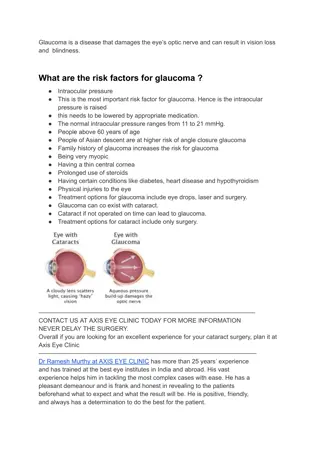
Glaucoma, the 3rd leading cause of blindness in Uganda, is primarily managed by General Ophthalmologists across 20 hospitals in the country. Uganda lacks Glaucoma subspecialists and specialized nurses. Equipment availability varies among hospitals. Cases in Uganda are typically identified through referrals, community outreach programs, routine eye checks, and walk-ins presenting symptoms like poor vision, pain, or the need for reading glasses. The common types of Glaucoma in Uganda include Congenital, Open Angle, Angle Closure, and Neovascular. Patients exhibit diverse presentations based on the Glaucoma type.

Download Presentation

Please find below an Image/Link to download the presentation.
The content on the website is provided AS IS for your information and personal use only. It may not be sold, licensed, or shared on other websites without obtaining consent from the author. Download presentation by click this link. If you encounter any issues during the download, it is possible that the publisher has removed the file from their server.
E N D
Presentation Transcript
SHARING BEST PRACTICES IN EYE CARE BETWEEN UGANDA AND THE UK A FOCUS ON GLAUCOMA BY: TINDYEBWA LUDOVICA K
AN OVERVIEW OF GLAUCOMA MANAGEMENT IN UGANDA. 1. Glaucoma is the 3rdcause of blindness in Uganda contributing approx. 6 %. In Uganda, Glaucoma is managed by General Ophthalmolgists who are distributed in 20 hospitals (10 Government, 10 private ) , 12 of which are located in Kampala. We also have OCOs who help us with screening both at District hospitals and in specialized eye clinics. They times initiate RX before referring to ophthalmologists. -We do not have Glaucoma subspecialists. - We do not have glaucoma specialized nurses. In terms of Equipment, most hospitals can do slitlamp biomicroscopy, applanation tonometry, a few hospitals use Pulse air and some still use schiotz tonometers. Very few hospitals have Visual field machines, OCT and fundus cameras. INTRODUCTION
Case finding Most patients walk in hospitals because of poor vision, loss of vision , pain or looking for reading glasses. Very few people are found during routine eye checks. Referrals from units run by OCOs and optometrists . Referrals from eye care community outreach programs . Referrals from fellow ophthalmologists.
2.TYPES OF GLUCOMA IN UGANDA and the common presentation. 1 . CONGENITAL GLAUCOMA ( both with and without anterior segment abnormalities ) These usually present in infancy . Pts are brought in by caretakers/ parents for a bupthalmos or cloudy corneas and photophobia. 2 . OPEN ANGLE GLAUCOMA : - Primary OAG: This is the commonest type of glaucoma we see. They present with poor vision, loss of vision or difficulties in reading . Some times found accidentally during fundoscopy. - Secondary OAG ( commonly due to trauma, uveitis, pseudoexfoliation and corticosteroids). Pts present with poor vision with or without painful red eyes depending on the cause. 3 .ANGLE CLOSURE GLAUCOMA : --Primary acute angle closure is rare. They come in with headache, severe eye pain and bLurring or rapid loss of vision. Eye exam findings are very high IOP, Corneal edema with mid dialated pupils and normal optic discs . - chronic angle closure is commoner and they present H/O of haloes around light and blurring of vision at night and early in the morning ( awake in dim light) and then diminishing vision. Usually you find optic disc cupping with normal IOP and Gonioscopy shows a shallow angle and areas of evidence of synechia --Secondary angle closure commonly to dislocated natural lens esp. after eye trauma . 4 . NEOVASCULAR GLAUCOMA usually associated with retinal neovascular diseases like PDR, and Retinal vein occlusions.
Case finding Walk ins to hospitals because of pain, poor , loss of vision or difficulties in reading or replacement of glasses. Cases found on routine fundoscopy during eye exams. Referrals by OCOs or medical ophthalmologists, optometrists. Screened cases from community outreach programmes. Babies are brought in by parents for abnormal eyes.
Patient management Diagnosis History- pt complaints sometimes give a clue to the type of glaucoma Exam :- visual acuity (VA) -Intraocular pressure (IOP). May or may not be raised in OAG, but always very high in acute angle closure. -slit lamp biomicroscopy for both anterior and posterior segments. Corneal edema is common in acute Angle closure and NVG due to very high IOP. -Optic Nerve disc changes are usually not present in acute angle glaucoma but disc cupping is diagnostic in OAG - Documentation of the optic nerve usually by vertical C:D > 0.5 ,ratio, cup depth, visible lamina cribrosa,notches, disc hemorrhages, bayonating vessels, and inequalities in the C:D of the two eyes.
ct Open angle glaucoma and chronic angle closure are diagnosed on optic nerve damage (cupping) Other glaucoma investigations/documentation methods now available in few facilities : -visual field tests in cases where the optic disc appearance is inconclusive but also to serve as a baseline for monitoring glaucoma progression. -OCT of the optic nerve. We take the vertical C: D. > 0.5. - Fundus photography A high IOP is a very important finding and may be the cause of the optic nerve damage. Gonioscopy to determine the angle characteristics and distinguish between OAG and Chronic angle closure.
Open Angle Glaucoma Treatment . The degree of optic damage (cupping ) determines the TARGET) IOP, which is presumed safe in minimizing further damage. As a guideline C:D 0.8 TO 1.0 , we target 15mmhg and less, C:D 0.6 to 0.7 target 18 and less, 0.5 and better 18-21 mmhg. Then go back to the IOP, because it s the only treatable factor. Depending on how high the IOP is , we choose the drugs / drug combinations that will push the IOP to the target.( using the IOP lowering effect, and C/Indications.) Surgery here is not attractive to patients, so we initially start with medical Rx , do cancelling , and finally most patients accept and get glaucoma surgery, especially those who fail to reach targeted IOP, defaulters on follow ups, those who find drugs expensive and co existing cataracts.
Currently available drugs are the : alpha agonists, beta adrenergic blockers, prostaglandin analogues and carbonic anhydrase inhibitors. GLAUCOMA SURGICAL PROCEDURE depend on the choice of the surgeon as per individual success rates. The commonest procedure here is a trabeculectomy with 5 FU or mitomycin C. Some people do them with multiple scleral flaps. Also combined procedure with cataract operations are commonly done. Some units are doing laser trabeculoplasty.
Treatment of angle closure Glaucoma Rx of Acute cases with high IOP depend on what is available to the Doctor. we IV mannitol if available to rapidly reduce the IOP. If not available, then Acetazolamide orally. When the IOP is reduced, we use 2 % pilocarpine eye drops to open the angle and later do a PERIPHERAL IRIDECTOMY ( YAG or surgical) depending on the facility. If the high IOP was due to lens displacement, then arrange for lens removal ( cataract surgery). If chronic angle closure without peripheral synechia, do PI, but if there is synechia, do trabeculectomy.
Treatment of Neovascular glaucoma Usually the IOP is very high We initially use Acetazolamide to reduce the IOP and topical antiglaucomas which suppress aqueous production. Where avastin is available , its given to reduce Rubeiosis and ultimately do a trabeculectomy or glaucoma drainage implant( few cases). Where possible PRP is done to induce regression of rubeiosis. The long term results of a trabeculectomy are not good esp. if rubeiosis is not handled.
Management of Congenital glaucoma We do Exam under General anaesthesia (EUA) Take IOP using schiotz or tonopens Measure corneal diameters Do fundoscopy Documentation. Post EUA, we control IOP with topical antiglaucoma , usually beta blockers and plan for glaucoma surgery. Common surgery here is trabeculectomy. Some people do goniotomy IOP monitoring is done regularly and for long . In some cases even after surgery antiglaucoma are ctd depending on the IOP AND at times, trabs are repeated.
CTD In summary Glaucoma management is controlling the Intraocular pressure to the level of minimizing or preventing visual loss. Although , there are general principles of management ,the clinician makes intervention decisions as per individual case. THANK YOU FOR YOUR PARTICIPATION.