Understanding Gram Staining in Bacterial Identification
Gram staining is a crucial technique in identifying bacterial organisms, developed by Hans Christian Gram. It involves differentiating bacteria into gram-positive and gram-negative groups based on cell wall properties. Gram staining has five basic steps and helps in determining bacterial characteristics for diagnostic and research purposes.

Download Presentation

Please find below an Image/Link to download the presentation.
The content on the website is provided AS IS for your information and personal use only. It may not be sold, licensed, or shared on other websites without obtaining consent from the author. Download presentation by click this link. If you encounter any issues during the download, it is possible that the publisher has removed the file from their server.
E N D
Presentation Transcript
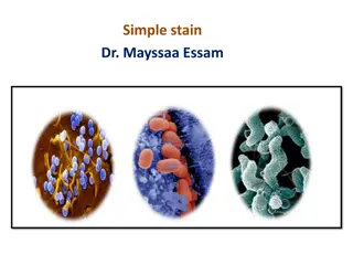
Gram staining The Gram staining is almost always the first step in the identification of a bacterial organism. The name comes from the Danish bacteriologist Hans Christian Gram, who developed the technique. Gram staining, also called Gram's method. It is a method of differentiating bacterial species into two large groups (gram-positive and gram-negative). While Gram staining is a valuable diagnostic tool in both clinical and research settings, not all bacteria can be definitively classified by this technique. For example Gram- indeterminate bacteria do not respond predictably to Gram staining; therefore, cannot be determined as either gram- positive or gram-negative. Gram-indeterminate bacteria are best stained using acid-fast staining techniques. Examples include many species of Mycobacterium, including M. tuberculosis and M. leprae.
There are five basic steps of the Gram staining: Fixation of bacteria onto a transparent surface (glass) by heat. Fixation is necessary so that the bacterial cells are not washed away by the treatment with subsequent solutions Application of primary stain (crystal violet, CV) to the heat-fixed smear of a bacterial culture. CV dissociates to form CV+ ion in the cell and interact with negatively charged components of the cells and cells become purple Addition of iodide, which binds to crystal violet and traps it in the cell by forming CV-I complex Decolorization with ethanol or acetone. A gram-negative cell loses its outer lipopolysaccharide membrane and the CV-I complex leave the cell through very thin peptidoglycan layer. In contrast, the thick peptidoglycan layer of gram-positive cell retain the purple color of CV-I. Counterstaining with safranin. After this stage gram negative cells become red or purple but gram positive remain violet
Gram staining Gram-positive bacteria are bacteria that give a positive result in the Gram staining test. Gram-positive bacteria take up the crystal violet stain used in the test, and then appear to be purple-colored when seen through a microscope. This is because the thick peptidoglycan layer in the bacterial cell wall retains the stain after it is washed away from the rest of the sample, in the decolorization stage of the test. Gram-negative bacteria cannot retain the violet stain after the decolorization step; alcohol used in this stage degrades the outer membrane of gram-negative cells making the cell wall more porous and incapable of retaining the crystal violet stain. Their peptidoglycan layer is much thinner and sandwiched between an inner cell membrane and a bacterial outer membrane, causing them to take up the counterstain (safranin) and appear red or pink.
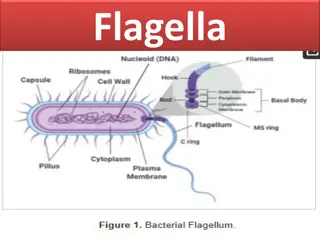
Functions of bacterial cell wall 1. Gives the cell a definite shape and structure and thus provides structural support 2. Protects against infection and mechanical stress 3. Separates interior of the cell from the outer environment 4. It enables transport of substances and information from the cell insides to the exterior and vice versa 5. Helps in osmotic-regulation 6. Prevents water loss 7. The physiological and biochemical activity of the cell wall helps in cell-cell communication 8. It prevents the cell from rupturing due to tugor pressure 9. Aids in diffusion of gases in and out of the cell 10.Also provides mechanical protection from insects and pathogens
Survival requirements of bacteria Chemical or nutritional requirements Physical or environmental requirements Carbon Nitrogen Sulfur Phosphorus Trace elements Oxygen Temperature pH Osmotic pressure
Temperature The minimum growth temperature: The lowest temperature at which the species are able to grow. The growth is seriously impaired below this temperature. The optimum growth temperature: The temperature at which the species grows best The maximum growth temperature: The highest temperature at which growth is possible and above which the growth is seriously impaired. Classification of bacteria based on their temperature requirements Psychrophiles Psychrotrophs Mesophiles Thermophiles Hyperthermophiles
Psychrophiles Psychrophiles or cryophiles are extremophilic organisms that are capable of growth and reproduction in cold temperatures, ranging from 10 C to +20 C. But their growth rate is highest around +100C. Exmp-Temperatures as low as 10 C are found in pockets of very salty water (brine) surrounded by sea ice. They are present in alpine and arctic soils, high- latitude and deep ocean waters, polar ice, glaciers and snowfields. Examples: Arthrobacter, Psycrobacter etc.
Psychrotrophs Psychrotrophic bacteria are bacteria that are capable of surviving or even thriving in a cold environment. They are found in soils, in surface and deep sea waters, in Antarctic ecosystems and in foods. They are responsible for spoiling refrigerated foods. Mesophile A mesophile is an organism that grows best in moderate temperature, neither too hot nor too cold, typically between 20 and 45 C. The habitats of these organisms include especially cheese, yogurt, and mesophile organisms are often included in the process of beer and wine making. Since normal human body temperature is 37 C, the majority of human pathogens are mesophiles.
Thermophile A thermophile is an organism, a type of extremophile, that thrives at relatively high temperatures, between 40 to 75 C. Many thermophiles are archaea. Thermophilic eubacteria are suggested to have been among the earliest bacteria. As a prerequisite for their survival, thermophiles contain enzymes that can function at high temperatures. Thermophiles are found in various geothermally heated regions of the Earth, such as hot springs, deep sea hydrothermal vents, as well as decaying plant matter, such as peat bogs and compost. Thermus aquaticus is a species of bacterium that can tolerate high temperatures. It is the source of the heat-resistant enzyme Taq DNA polymerase, one of the most important enzymes in molecular biology because of its use in the polymerase chain reaction (PCR) DNA amplification technique.
Hyperthermophile: A hyperthermophile is an organism that thrives in extremely hot environments: from 65 C upwards. An optimal temperature hyperthermophiles is above 80 C. Hyperthermophiles are a subset of extremophiles, micro- organisms within the domain Archaea, although some bacteria are able to tolerate temperatures of around 100 C (212 F). Some bacteria can live at temperatures higher than 100 C at very deep sea where water does not boil because of high pressure. for the existence of Example: Methanopyrus kandleri can survive and reproduce at 122 C.
pH Most bacteria grow best in a narrow pH range near neutrality, between pH 6.5 and 7.5 Very few bacteria grow at an acidic pH below about pH 4. This kind of bacteria are known as acidophiles. This is why a number of foods, such as pickles, and many cheeses, are preserved from spoilage by acids produced by bacterial fermentation. When bacteria are cultured in the laboratory, they often produce acids that eventually interfere with their own growth. To neutralize the acids and maintain the proper pH, chemical buffers are included in the growth medium.
Osmotic pressure Microorganisms obtain almost all their nutrients in solution from the surrounding water Plasmolysis: High osmotic pressures have the effect of removing necessary water from a cell. When a microbial cell is in a solution whose concentration of solutes is higher than in the cell, the cellular water passes out through the plasma membrane to the high solute concentration, a phenomenon termed as plasmolysis. Salted fish, honey, and sweetened condensed milk are preserved largely by the addition of high salt/sugar; the high salt or sugar concentrations increase osmotic pressure which draw water out of any microbial cells that are present and thus prevent their growth Deplasmolysis: Conversely when a bacterial cell is placed in a solution having lower concentration than the cell, water from the outside will enter into the cell by osmosis which will cause the increase of osmotic pressure inside the cell and if the pressure is very high the cell will burst. This phenomenon is called deplasmolyisis.