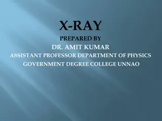
Understanding Radiographic Testing: History, Science, and Radiation Safety
Exploring the history and science behind Radiographic Testing (RT), from the discovery of X-rays to the development of methods using gamma radiation. Learn about the terminology, natural radiation, and the characteristics of radiation in various forms. Understand the importance of safety measures in utilizing X-rays and gamma rays for non-destructive testing.

Download Presentation

Please find below an Image/Link to download the presentation.
The content on the website is provided AS IS for your information and personal use only. It may not be sold, licensed, or shared on other websites without obtaining consent from the author. Download presentation by click this link. If you encounter any issues during the download, it is possible that the publisher has removed the file from their server.
E N D
Presentation Transcript
Radiographic Testing Topics History of RT Science of Radiation Radiation Terminology Radiation Safety
Radiographic Testing Radiographic Testing (RT) Definition: An NDT method that utilizes x-rays or gamma radiation to detect discontinuities in materials, and to present their images on recording medium. History X-Rays X-rays discovered in 1895 by Wilhelm Roentgen 1896 used by physicians to locate bullets in soldiers 1913 development of high vacuum x-ray tubes enabled voltages of 100,000 V. Increased penetrating power of x-rays led to industrial applications 1931 GE developed 1,000,000 V x-ray generator ASME approved use of x-ray to test fusion-welded pressure vessels
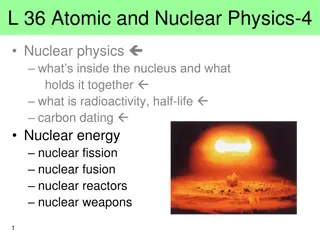
Radiographic Testing History Natural Radiation 1896 Henri Becquerel discovers and identified uranium as radioactive material 1898 Pierre and Marie Curie discovered polonium, followed by radium ( shining element) Radium capable of filming through 10-12 thick steel castings Used extensively during WWII as part of U.S. Navy shipbuilding program 1946 cobalt and iridium became available Both stronger and cheaper than radium
Radiographic Testing What is radiation? In the simplest sense, radiation is the emission of energy as electromagnetic waves. Below is a graphic of the various forms of radiation present in our environment.( Also known as the Electromagnetic Spectrum)
Radiographic Testing What is radiation? As seen in the previous graphic the wavelength and frequency of the radiation describes what form it takes. Radiation is in the form of radio Waves to visible light to gamma rays. The higher the frequency the more energetic and penetrating the radiation. This high energy radiation is generally classified as a X-ray or gamma ray and is characterized by its ability to ionize atoms that are exposed to it, thus called ionizing radiation.
Radiographic Testing What is radiation? In the simplest sense, radiation is the emission of energy as electromagnetic waves. X-rays and gamma rays are types of electromagnetic radiation of shorter wavelengths than visible light: visible = 600 Angstroms, gamma rays = 0.0001 A shorter wavelengths permit penetration through materials high energy levels break chemical bonds *Leads to destruction of living tissue X-rays and gamma rays differ only in source of origin. X-rays come from the electron shell and gamma rays come from the Nucleus of a Atom. x-rays = 1.0 A,
Radiographic Testing X-ray or Gamma Ray radiation Properties Undetectable by human senses Cannot be seen, felt, heard, or smelled Possesses no charge or mass Referred to as photons (packets of energy) Generally travels in straight lines at the speed of light (can bend at material interfaces) Characterized by frequency, wavelength, and velocity Part of electromagnetic spectrum but not influenced by electrical or magnetic fields They have enough energy to ionize matter
Radiographic Testing Atoms Atom - the smallest particle of any element that retains the characteristics of that element Subatomic particles protons, electrons, neutrons
Radiographic Testing Atoms (continued) Nucleus of atom contains the positively-charged protons and neutrally-charged neutrons Electrons negatively-charged particles that travel at high speed around the nucleus Atomic number based on an element s number of protons Ex. Carbon (C) has atomic number 6, Potassium (K) has atomic number 19 Isotopes atoms of the same element with different number of neutrons Atomic mass approximately equal to sum of protons and neutrons
Radiographic Testing Radioactivity Defined as the release of energy and matter that results from changes in the nucleus of an atom An atom that contains additional neutrons is unstable In order to move to a more stable state, the nucleus seeks to remove the extra neutrons, thereby emitting radiation Atoms with atomic numbers > 83 referred to as radioisotopes they have unstable nuclei and are radioactive
Radiographic Testing Radioactive Decay Defined as the spontaneous breakdown of an atomic nucleus resulting in the release of energy and matter from the nucleus Occurs by: Alpha decay (emits 2 protons and 2 neutrons from the nucleus) Beta decay (a neutron split into a proton and an electron) Gamma decay (energy in the form of gamma radiation emitted from the nucleus) Alpha and beta decay involve particles Example nuclear reaction: 92U238 90Th234+ 2He4+ gamma rays
Radiographic Testing Radioactive Decay (continued) When an atom undergoes radiographic decay, it emits one or more forms of radiation with sufficient energy to ionize the atoms with which it interacts Ionizing radiation high speed subatomic particles ejected from the nucleus, or gamma rays emitted by the either the nucleus or orbital electrons Ionization complete removal of an electron from an atom following the transfer of energy from a passing charged particle
Radiographic Testing Radioactive Decay (continued) Activity quantity which expresses the degree of radioactivity or the radiation-producing potential of a given amount of radioactive material Given in units of Curie (English system) or Becquerel (SI) One Curie is quantity of radioactive material in which 2.22 * 1012disintegrations per minute or 3.7 * 1010atoms disintegrate per second (move to a more stable state) 1 Becquerel = quantity of radioactive material in which 1 atom disintegrates per second 1 Curie = 3.7 * 1010Becquerel Specific activity activity per unit mass or volume
Radiographic Testing Half-Life Defined as the time required for the activity of a particular radioisotope to decrease to half of its original value Varies for different radioisotopes Ranges from microseconds to billions of years (uranium) Half-life of Cobalt-60 = 5.3 years Half-life Iridium-192 = 74 days Carbon-14 dating used to approximate the age of fossils Decays with a half-life of 5730 years
Radiographic Testing Half-Life Example Example: You originally have a 30-Curie source of Cobalt-60. 1. How many Curies will you have in 5.3 years? Answer: The source will have decayed by one half-life in 5.3 years, therefore half of the original source will remain, or 15 Curies. 2. How long will it take for the source to decay to 3.75 Curies? Answer: Every 5.3 years the source will be reduced by half. In 1 half-life, the source will be reduced to 15 Curies. In 2 half-lives, the source will be down to 7.5 Curies. Finally, in 3 half-lives, the source will be 3.75 Curies. Therefore, it will take a total of 3 half-lives (or 15.9 years) for the source to decay to 3.75 Curies.
Radiographic Testing Penetrating Power of Radiation Only x-rays and gamma rays can penetrate solid materials to a significant extent (alpha and beta particles can penetrate skin only up to 8 mm) Transmission of radiation depends on: Material thickness Material density Energy of the photons Photons that are not transmitted are attenuated (absorbed or scattered)
Radiographic Testing Radiation Safety Science of radiation protection (also called health physics) developed to address acceptable radiation levels to reduce risk of injury to humans Primary health concern (for chronic, not acute exposure) is increased risk for cancer Depends on amount of radiation dose, duration of dose, and specific body parts exposed Possession and use of radioactive materials strictly regulated by the Nuclear Regulatory Commission (NRC) as specified in Title 10 of the Code of Federal Regulations (CFR) 20
Radiographic Testing Radiation Safety (continued) By 1900 it was understood that use of x-rays and gamma radiation required safety precautions to protect one s health 1920 s routine use of film badges for personnel monitoring begun, genetic effects of radiation recognized Origin of radiation Natural (sun, radon gas) Man-made (x-rays, gamma rays)
Radiographic Testing Radiation Energy Different radioactive materials and x-ray generators produce radiation at different energies Energy of radiation responsible for the ability to penetrate matter Measured in electronvolts (eV) or kiloelectronvolts (keV) An electronvolt is the energy gained by an electron passing through a potential difference of 1 volt Energy adjustable on an x-ray generator Energy an unchangeable characteristic of a radioisotope
Radiographic Testing Radiation Intensity Amount of energy passing through a given area (perpendicular to the direction of radiation travel) in a given unit of time Survey meter used to measure intensity or radiation dose rate Units in mRem/hr. or Rem/hr. Frequently referred to as the most important tool a radiographer has to determine the presence and intensity of radiation Per the inverse square law, the intensity varies inversely with the square of the distance
Radiographic Testing Dose Exposure measure of the strength of a radiation field Units of Roentgen or R Dose (also called absorbed dose) amount of ionizing energy absorbed by an object Units of Rad ( radiation absorbed dose ) Dose equivalent relates absorbed dose to biological effect of dose equals absorbed dose times a quality factor (quality factor = 1 for x-ray and gamma radiation in humans) Units of Rem ( Roentgen equivalent in man ) For humans, 1 R = 1 Rad = 1 Rem
Radiographic Testing Dose vs. Dose Rate Dose rate measure of how fast a radiation dose is being received dose rate = dose / time Units of R/hr, mR/hr, Rem/hr, mRem/hr (same units as intensity) To calculate a person s dose, multiply the dose rate (measured with a survey meter) by the duration of exposure dose = dose rate * time Example: If the radiation intensity in a particular area is 5 mRem/hr, and the person remains in that area for 30 minutes, what is the person s radiation dose? dose = 5 mRem/hr * 0.5 hr = 2.5 mRem
Radiographic Testing Biological Effects of Radiation Absorbed dose depends on: Intensity of radiation source, Distance from source, Time of exposure Ionization of living tissue causes molecules in cells to be broken apart Biological effects vary with: Type of radiation (penetrating ability) Size of dose received Dose rate Body part exposed (extremities can handle more exposure than blood-forming organs) Age of individual (cell divisions slow with age older people less sensitive to ionizing radiation)
Radiographic Testing Stochastic and Nonstochastic Effects Stochastic effects are delayed and difficult to specifically correlate to radiation exposure (cancer) Nonstochastic effects are acute effects Directly proportional to size of dose Symptoms Skin reddening Skin and tissue burns Cataract formation Sterility Radiation sickness (nausea, vomiting, diarrhea, headache, fever, dizziness, weakness, hair loss) Death
Radiographic Testing Important NRC Codes 10CFR19 Notices, Instructions, and Reports to Workers: Inspection and Investigations 10CFR20 Standards for Protection Against Radiation 10CFR34 Licenses for Industrial Radiography and Radiation Safety Requirements for Industrial Radiographic Operations Guidelines established to both prevent acute exposure and limit chronic exposure to acceptable levels Desire to maintain all exposure as low as reasonably achievable referred to as ALARA
Radiographic Testing Exposure Limits Effect of exposure (dose received in a 24-hour period): 0-25 Rem no evident injury (blood changes detectable at 5 Rem) 400-500 Rem results in death in 50% of cases 1000+ Rem results in death in ALL cases Annual Occupational Limits Total effective dose 5 Rem For Declared pregnant women - 0.5 Rem during gestation period For people younger than age 18 0.5 Rem
Radiographic Testing Exposure Limits Continued Annual Non-Occupational Limits (for general public exposure to industrial ionizing radiation) 0.1 Rem (2% of occupational limit) Doesn t include natural background radiation (0.3 Rem) or radiation from man-made sources, such as medical x-rays (0.05 Rem)
Radiographic Testing Controlling Radiation Exposure Remember: TIME, DISTANCE, and SHIELDING Recall that dose = dose rate * time Collimator device used to direct radiation onto a part and shield the radiation in other directions In general, the denser the material the greater the shielding Most effective shielding by depleted uranium used to shield the source in gamma ray cameras Lead and concrete commonly used for shielding
Radiographic Testing Controlling Radiation Exposure Recall inverse square law the radiation intensity varies inversely with the square of the distance I1*D12= I2*D22 Example: If the intensity at 1 from the source is 500 Rem/hr, at what distance is the intensity 2 mRem/hr? Solution: Let I1= 500 Rem/hr; D1= 1 foot; I2= 2 mRem/hr Solve for D2. D2= Square root (I1*D12/ I2) = 500 feet Signs must be posted at intensity boundaries (usually 2 mRem/hr and 100 mRem/hr) to warn individuals that radiographic testing is in progress
Radiographic Testing Half-Value Layer Defined as the thickness of a material needed to reduce the radiation intensity to of its original value To provide shielding Depends on radioisotope used for gamma radiation (or voltage if radiation source is an x-ray) Depends on voltage if radiation source if an x-ray
Radiographic Testing Radiation Safety Officer Radiation Safety Officer (RSO) - individual authorized by company to ensure radiation activities performed safely and per approved procedures and regulations Radiographers required to wear personnel monitoring devices to aid in tracking and minimizing their radiation exposure
Radiographic Testing Personnel Monitoring Devices Pocket dosimeter Similar in appearance to small marker Reads radiation dose (not dose rate) Allows workers to track daily dose received (reset at the start of each shift) Audible alarm rate meter Sounds an alarm at a preset dose rate (typically 500 mRem/hr) NOT a replacement for a survey meter!
Radiographic Testing Personnel Monitoring Devices Pocket dosimeter
Radiographic Testing Personnel Monitoring Devices (continued) Film badge Piece of radiation-sensitive film that provides a permanent record of radiation dose received TLD (thermoluminescent dosimeter) Often used instead of a film badge (may be reused) Both film badges and TLDs typically worn for 1-3 months before being processed to determine dose OSL (Optically Stimulated Luminescent) dosimeter Often used instead of a TLD or film badge (may be reused) Can be work for longer periods of time due to good fading.
Radiographic Testing Personnel Monitoring Devices (continued) Various monitoring devices.
Radiographic Testing Radiography Equipment X-Ray Generators X-rays are generated by directing a stream of high-speed electrons at a target material, such as tungsten, which has a high atomic number Interaction with the tungsten atoms slows or stops the electrons, and x-rays are produced Major components of an x-ray machine Tube contains a cathode (coiled wire) and an anode (target) operates in a vacuum High voltage generator Control console (to adjust output) Cooling system
Radiographic Testing X-Ray Generators (continued) X-ray control console Increasing current (or milliamperage) increases number of electrons that flow from the cathode to the anode thus increases the x-ray intensity Increasing the voltage increases the speed at which the electrons travel thus increasing the energy or penetrating power of the x-ray
Radiographic Testing Radiography Equipment Gamma Sources Man-made radioactive sources produced by introducing an extra neutron to atoms of the source material by neutron bombardment As the atoms shed the neutron energy is released in the form of gamma ray Co-60 and Ir-192 common industrial gamma ray sources due to: high energies Portability (as opposed to x-ray machines) Disadvantage of gamma sources cannot be turned off, therefore source must be isolated and shielded within an exposure device (camera)
Radiographic Testing Radiography Equipment Gamma Sources Sources typically consist of multiple pellets loaded into a stainless steel capsule to obtain desired activity level (number of Curies) Capsule sealed by welding Attached to a short flexible cable called a pigtail Housed in a shielding device referred to as an exposure device or camera Activity of source governs amount of shielding required Co-60 has a higher comparable energy (1.25 MeV) than Ir- 192 (460 keV) Co-60 cameras typically weigh 500 lbs vs. ~45 lbs for Ir-192 cameras
Radiographic Testing Radiography Equipment Gamma Sources
Radiographic Testing Radiography Equipment Gamma Sources
Radiographic Testing Radiography Equipment Exposure Vaults & Cabinets Exposure vaults and cabinets allow personnel to work safely in the area while exposures are taking place. Exposure vaults tend to be larger walk in rooms with shielding provided by high-density concrete block and lead. Exposure cabinets are often self-contained units with integrated x-ray equipment and are typically shielded with steel and lead to absorb x-ray radiation. They are equipped with protective interlocks that disable the system if anything interrupts the integrity of the enclosure.
Radiographic Testing Performing Radiography Exposure device connected to source tube on one end and a crank- out mechanism on the other end Source tube is positioned as needed to obtain radiograph of component (with the source itself safely housed in the exposure device) From as far away as possible, the radiograph turns the crank to move the source from the camera to the tip of the source tube After the proper exposure time (calculated based on source strength, source to film distance, and thickness of the component), the radiographer cranks the source back into the camera The radiographer must use the survey meter to confirm the source fully retracted back into the camera
Radiographic Testing Radiograph Image Properties Sensitivity a measure of the quality of the image in terms of the smallest detail that may be detected Image Quality Indicators (IQIs or penetrameters) devices used to indicate the quality level or sensitivity of the radiograph (plaque type or wire type) Depends on contrast and definition Contrast degree of density difference between two areas on a radiograph Definition degree of sharpness of the radiographic image Codes require a minimum unsharpness
Radiographic Testing Radiograph Image Properties Density degree of film darkening If 0.01% of transmitted light reaches far side of film the density is 4.0 (based on an exponential equation) If 1.0% of transmitted light reaches far side of film the density is 2.0 Many governing codes require densities of 2-4% (don t want image too light or too dark)
Radiographic Testing Radiograph Image Properties Densitometer device used to measure film density Density through the IQI must meet code (2-4%) Also, the density in the area of interest (the weld perhaps), must satisfy plus/minus criteria Darkest area must have a density of not more than 30% of the density under the IQI Lightest area must have a density of not less than 15% of the density under the IQI