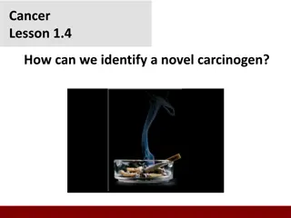
Understanding Therapeutic Strategies Targeting Apoptosis in Cancer

Apoptosis, a form of programmed cell death, is crucial for cellular homeostasis. In cancer, inhibition of apoptosis processes allows cancer cells to survive and proliferate abnormally. Developing targeted treatments for apoptosis pathways is essential for improved outcomes in cancer therapy. Cell death mechanisms, including apoptosis, play vital roles in growth, development, and tissue maintenance. Different strategies are being explored to enhance the use of pro-apoptotic agents. Apoptosis can be triggered by various internal and external factors, highlighting its importance in cancer treatment.

Download Presentation

Please find below an Image/Link to download the presentation.
The content on the website is provided AS IS for your information and personal use only. It may not be sold, licensed, or shared on other websites without obtaining consent from the author. Download presentation by click this link. If you encounter any issues during the download, it is possible that the publisher has removed the file from their server.
E N D
Presentation Transcript
Therapeutic strategies: Targeting apoptosis in cancer Last updated: September 2020
Introduction Apoptosis, a form of programmed cell death, is a key aspect of cellular homeostasis1 In cancer, the processes and signals that promote apoptosis are inhibited, allowing cancer cells to survive and proliferate in a dysregulated manner2,3 A deeper understanding of cell death pathways in cancer is essential for the development of precisely targeted treatments or synergistic treatment combinations4 A number of different strategies are under investigation to optimise outcomes with pro-apoptotic agents3 1. Fuchs Y, Stellar H. Cell 2011;147(4):742 58; 2. Sharma A, et al. Cancers 2019;11:1144; 3. Jan R, Chaudry G. Adv Pharm Bull 2019;9(2):205 18; 4. Ricci MS, Zong WX. Oncologist 2006;11(4):342 57.
Cell death is a necessary biological function involved in growth, homeostasis and maintenance of healthy cells Programmed cell death plays a fundamental role in animal development and tissue homeostasis1 The cell death mechanism is involved during:1,2 Arrangement of cells during morphogenesis Regulation of cell number during homeostasis Elimination of abnormal cells during pathogenesis 1. Fuchs Y, Steller H. Cell 2011;147(4):742 58; 2. Jan R, Chaudhry G. Adv Pharm Bull 2019;9:205 18.
Cell death typically occurs as a result of either necrosis or a programmed cell death such as apoptosis* Necrosis occurs when cells die in response to injury or cellular stress, by swelling and rupturing1,2 Apoptosis occurs when cells die during (e.g.) homeostasis, by condensation without losing membrane integrity - and removal by phagocytosis1,2 Injured cell Cell swells Cell and chromatin condense and shrink Cell becomes leaky (blebbing) Cell buds into apoptotic bodies Cell breaks down Apoptotic bodies removed by phagocytosis Redundant cell *Apoptosis is one example of programmed cell death; other examples include autophagy and necroptosis.2 Figures adapted from Van Cruchton S, Van den Broeck W. 2002.3 1. Jacobson MD, et al. Cell 1997;88(3):347 54; 2. Jan R, Chaudhry G. Adv Pharm Bull 2019;9:205-18; 3. Van Cruchton S, Van den Broeck W. Anat Histol Embryol 2002;31:214 23.
Apoptosis can be triggered by a range of internal and external circumstances1,2 Hormone-dependent changes in tissues OH Cell loss in proliferating populations H Normal development H H HO Elimination of lymphocytes Response to radiation or Apoptosis chemotherapeutic drugs Elimination of abnormal cells 1. Fuchs Y, Steller H. Cell 2011;147(4):742 58; 2. Elmore S. Toxicol Pathol 2007;35(4):495 516.
Apoptotic triggers act through the intrinsic or extrinsic signalling pathways to cause apoptosis The intrinsic pathway is initiated by apoptogenic triggers or cellular stress1 This leads to the insertion of the Bcl-2 proteins, Bax/Bak, into the mitochondria, followed by cytochrome C release2 Cytochrome C combines with Apaf-1 and procaspase-9 to form an apoptosome, which activates the caspase cascade to terminate the cell3 6 SMAC, a pro-apoptogenic mitochondrial protein, is also released into the cytosol, where it antagonizes IAPs to allow caspase activation and subsequent apoptosis5 BH3 protein Bax/Bak Mitochondrion Cytochrome C Apaf-1 Procaspase-9 SMAC Apoptosome XIAP IAP Caspase 3,6,7 Apoptosis Apaf, apoptotic protease activating factor; Bak, Bcl-2 antagonist/killer; Bax, Bcl-2-associated X protein; Bcl, B-cell lymphoma; IAP, inhibitor of apoptosis proteins; SMAC, second mitochondrial- derived activator of caspases (also known as DIABLO, direct IAP-binding protein with low pI); XIAP, X-linked inhibitor of apoptosis protein. Figure adapted from Baig et al. 2016.6 1. Green DR, Llambi F. Cold Spring Harb Perspect Biol 2015;7:a006080; 2. Zhang M, et al. Sci Rep 2017;7:2635; 3. Bratton SB, Salveson GS. J Cell Sci 2010;123(19):3209 14; 4. Jan R, Chaudhry G. Adv Pharm Bull 2019;9:205 18; 5. Martinez-Ruiz G, et al. J Exp Clin Cancer Res 2008;27:48; 6. Baig S, et al. Cell Death Dis 2016;7:e2058.
Apoptotic triggers act through the intrinsic or extrinsic signalling pathways to cause apoptosis Activated death receptor The extrinsic pathway is triggered by external stimuli or ligand molecules such as TRAIL1 Activation of the extrinsic pathway then triggers the caspase cascade to promote apoptosis1 DISC Caspase 8 Procaspase 3,6,7 Caspase 3,6,7 Apoptosis DISC, death-inducing signaling complex; TRAIL, tumor necrosis factor-related apoptosis-inducing ligand. Figure adapted from Baig et al. 2016.2 1. Green DR, Llambi F. Cold Spring Harb Perspect Biol 2015;7:a006080; 2. Baig S, et al. Cell Death Dis 2016;7:e2058.
Evasion of apoptotic processes in cancer
Cancer cells survive and proliferate via evasion of apoptogenic triggers Hormone-dependent changes in tissues OH Cell loss in proliferating populations Abnormal regulation of programmed cell death is associated with many diseases, including cancer1 H Normal development H H HO Defects in DNA repair and chromosome segregation normally trigger cell death to remove these genetically unstable cells2 Defects in apoptosis: Allow genetically unstable cells to survive and allow selection of progressively aggressive clones2,3 Allow neoplastic cells to survive beyond their typical lifespan, providing protection from hypoxia and oxidative stress as the tumor mass expands3 Allow epithelial cells to survive in a suspended state, detached from the extracellular matrix, which facilitates metastasis3 Elimination of lymphocytes Response to radiation or Apoptosis chemotherapeutic drugs Elimination of abnormal cells 1. Fuchs Y, Stellar H. Cell 2011;147(4):742 58; 2. Hassan M, et al. Biomed Res Int 2014;2014:150845; 3. Reed JC. Cancer Cell. 2003;3(1):17 22.
Cancer cells inhibit the intrinsic pathway by upregulating anti-apoptotic Bcl-2 proteins BH3 protein Stimulus, e.g. ROS Bcl-2 proteins all of whom contain BH1-4 domains are key regulators of the intrinsic pathway1,2 Release of activators (e.g.) PUMA Bax/Bak The intrinsic pathway is initiated by release of pro-apoptotic BH3 activators (i.e. BIM, PUMA, tBID) that activate multi-domain effectors (e.g. Bax; Bak) to promote MOMP2 Activation of apoptotic pathway via MOMP Mitochondrion Cytochrome C Apaf-1 Procaspase-9 SMAC Apoptosome XIAP IAP Cancer cells often express elevated levels of pro-apoptotic BH3-only proteins3 Thus, cancer cells will typically upregulate anti- apoptotic Bcl-2 proteins in order to sequester unbound pro-apoptotic proteins, preventing MOMP and activation of the apoptotic cascade3 Caspase 3,6,7 Apoptosis Bak, Bcl-2 antagonist/killer; Bax, Bcl-2 associated X protein; Bcl, B-cell lymphoma; BH, Bcl-2 homology; BIM, Bcl-2 interacting mediator of cell death; MOMP, mitochondrial outer membrane permeabilization; PUMA, p53-upregulated modulator of apoptosis; tBID, truncated BH3 interacting domain death agonist. Figure adapted from Baig et al. 2016.4 1. van Delft MF, Huang DCS. Cell Res 2006;16:203 13; Happo L, et al. J Cell Sci 2012;125(5):1081 7; 3. Sharma A, et al. Cancers 2019;11:1144; 4. Baig S, et al. Cell Death Dis 2016;7:e2058.
Cancer cells inhibit the intrinsic pathway by suppressing p53 expression The p53 tumor suppressor protein upregulates pro-apoptotic BH3-only proteins such as PUMA and NOXA1 Tumor cell3,4 The p53 gene is mutated or deleted in many human cancers, inactivating its suppressor activity2 In some cancers with wild-type p53 status, its function is effectively inhibited by amplified expression of MDM2, which is the primary cellular inhibitor of p531,2 Thus, by inhibiting or deleting p53 activity, malignant cells remove a key driver for apoptosis and maintain their proliferative state BH, Bcl-2 homology; MDM2, murine double minute 2; PUMA, p53 upregulated modulator of apoptosis. 1. Aubrey BJ, et al. Cell Death Differ 2018;25:104 13; 2, Shangary S, Wang S. Clin Cancer Res 2008;14(17):5318 24. 3. Nag S, et al. J Biomed Res 2013;27(4);254 71; 4. Zhao Y, et al. Acta Biochim Biophys Sin 2014;46:180 9.
Cancer cells inhibit the intrinsic pathway by modifying cell metabolism Tumor cell Specific Bcl-2 proteins can be regulated by metabolite stresses1 Glucose deprivation may induce PUMA via p53 induction1 p53 inhibits glycolysis through down-regulation of glucose transporters, glycolytic enzymes, and inhibition of hypoxic-inducible factors1 p53 Oncogene Glycolytic environment Activation of oncogenes (e.g. RAS, AKT, MYC) as well as loss of genes such as p53 drive aerobic glycolysis in cancer cells and promote a glycolytic phenotype,1,2 thereby subverting micronutrient-driven apoptotic activation Bcl, B-cell lymphoma; PUMA, p53-upregulated modulator of apoptosis; tBID, truncated BH3 interacting domain death agonist. 1. Sharma A, et al. Cancers 2019;11:1144; 2. Zheng J. Oncol Lett 2012;4:1151 7.
Cancer cells inhibit the intrinsic pathway by offsetting oxidative stress mechanisms Higher ROS can upregulate cell death pathways1 Low ROS may upregulate proliferative pathways and, thus, promote tumorigenesis2 Decreased ROS = increased tumorigenesis Due to their high metabolic activity, cancer cells often contain increased ROS2,3 Consequently a cancer cell must synthesize antioxidants in order to retain the cell s redox homeostasis2,3 Increased ROS = increased cell death activation Cancer cells may also increase the uptake of antioxidant nutrients and metabolic enzymes2 to moderate intracellular ROS ROS, reactive oxygen species. 1. Redza-Dutordoir M, et al. Biochim Biophys Acta 2016;1863:2977 92; 2. Sharma A, et al. Cancers 2019;11:1144; 3. Liou GY, Storz P. Free Radic Res 2010;44(5):479 96.
Cancer cells have evolved strategies to evade extrinsic pathway-induced apoptosis Activated death receptor The extrinsic pathway is mediated by the death receptors TNFR1, CD95, FasR, APO-1, DR4 and DR51 DISC Death receptors are activated by regulatory ligands such as TRAIL, which induce DISC formation and initiate the apoptotic pathway1 Caspase 8 Procaspase-3,6,7 In cancer cells, TRAIL resistance may be caused by various genetic mutations (resulting in altered apoptotic signaling proteins), or overexpression of anti-apoptotic Bcl-2 proteins1 Caspase 3,6,7 Apoptosis APO-1, apoptosis antigen 1; Bcl, B-cell lymphoma; CD95, cluster of differentiation 95; DISC, death-inducing signaling complex; DR, death receptor; FasR, Fas receptor; TNFR1, tumor necrosis factor receptor 1; TRAIL, TNF-related apoptosis-inducing ligand. Figure adapted from Baig et al. 2016.2 1. Ukrainskaya VM, et al. Acta Naturae 2017;9(3):55 63; 2. Baig S, et al. Cell Death Dis 2016;7:e2058.
Targeted induction of apoptosis in cancer cells A deeper understanding of cell death pathways in cancer is essential for the development of precisely targeted treatments or synergistic treatment combinations1* DR ligands2 Attenuation of Bcl-2 proteins2 BH3 protein DISC p53/MDM2 expression2 Bax/bak Caspase 8 Mitochondrion Cytochrome C Apaf-1 Procaspase-3,6,7 Procaspase-9 SMAC SMAC expression2 Caspase 3,6,7 Apoptosome XIAP IAP Caspase 3,6,7 Apoptosis Apoptosis Apaf, apoptotic protease activating factor; Bak, Bcl-2 antagonist/killer; Bax, Bcl-2-associated X protein; Bcl, B-cell lymphoma; DISC, death-inducing signaling complex; DR, death receptor; IAP, inhibitor of apoptosis proteins; MDM2, murine double minute 2; SMAC, second mitochondrial-derived activator of caspases (also known as DIABLO, direct IAP-binding protein with low pI); X-linked inhibitor of apoptosis protein. *These are investigational compounds and have not been approved. Their efficacy and safety have not been established. Figures adapted from Baig et al. 2016.3 1. Ricci MS, Zong WX. Oncologist 2006;11(4):342 57; 2. Jan R, Chaudry G. Adv Pharm Bull 2019;9(2):205 18; 3. Baig S, et al. Cell Death Dis 2016;7:e2058.
Approaches to target the intrinsic pathway: Bcl-2 attenuation Bcl-2 inhibitors1,2* Antisense oligonucleotides* to enhance sensitivity to cytotoxic drugs3,4 Strategies used to inhibit the anti-apoptotic Bcl-2 family include: Cyclin-dependent kinase inhibitors* to downregulate expression of factors such as Bcl-2, Mcl-1 and XIAP5 BH3-mimicking agents6,7* Bcl, B-cell lymphoma; BH, Bcl-2 homology; Mcl-1. myeloid cell leukemia-1; XIAP, X-linked inhibitor of apoptosis protein. *These are investigational compounds and have not been approved. Their efficacy and safety have not been established. 1. NCT02427451. Available at: https://clinicaltrials.gov/ct2/show/NCT02427451. Accessed July 2020; 2. NCT04277637. Available at: https://clinicaltrials.gov/ct2/show/NCT04277637. Accessed July 2020; 3. Baig S, et al. Cell Death Dis 2016;7:e2058; 4. Lu AQ, et al. Int J Clin Exp Med 2018;11(7):6767 75; 5. Boffo S, et al. J Exp Clin Cancer Res 2018;37:36; 6. Jan R, Chaudry G. Adv Pharm Bull 2019;9(2):205 18; 7. Mukherjee N, et al. Cell Death Dis 2018;9:907.
Spotlight on p53/MDM2 Tumor cell4 6 The tumor suppressor gene p53 impacts both cell cycle arrest and apoptosis1 Interaction inhibitor* p53 activity is inhibited by MDM2 upregulation Antisense oligonucleotides and MDM2 inhibitors* have been developed to reduce MDM2 overexpression and trigger wild-type p53 activity1,2 Another approach uses small molecules that reactivate wild-type function* of mutant p531,3 MDM2, mouse double-minute 2; wt, wild-type. *These are investigational compounds and have not been approved. Their efficacy and safety have not been established. 1. Jan R, Chaudry G. Adv Pharm Bull 2019;9(2):205 18; 2. Rayburn ER, et al. Anticancer Agents Med Chem 2009;9(8):882 903; 3. Di Agostino S, et al. J Exp Clin Cancer Res 2019;38(1):290; 4. Rudolph D, et al. Presented at the Annual Meeting of the American Association for Cancer Research 2018. Abstract 4868 and poster; 5. Nag S, et al. J Biomed Res 2013;27(4);254 71; 6. Zhao Y, et al. Acta Biochim Biophys Sin 2014;46:180 9.
Spotlight on SMAC SMAC is released in response to Bcl-2 activity on the mitochondrion, where it binds to and induces IAP degradation to promote apoptosis1 In cancer cells, elevated IAP expression increases cell survival, tumor growth and metastasis1 BH3 protein Bax/bak SMAC mimetics* bind to cellular IAPs to restore effector caspase function and are under investigation alone or as combination therapy2,3 Mitochondrion Cytochrome C Apaf-1 Procaspase-9 SMAC Apoptosome XIAP IAP However, SMAC is overexpressed in some cancers, suggesting a role in non-apoptotic cancer cell survival and a possible role for reducing SMAC expression in some cancers4,5 Caspase 3,6,7 Apoptosis Bcl, B-cell lymphoma; IAP, inhibitor of apoptosis protein; SMAC, second mitochondrial-derived activator of caspases (also known as DIABLO, direct IAP-binding protein with low pI). *These are investigational compounds and have not been approved. Their efficacy and safety have not been established. Figure adapted from Baig et al. 2016.6 1. Bai L, et al. Pharmacol Ther 2014;144(1):82 95; 2. Jan R, Chaudry G. Adv Pharm Bull 2019;9(2):205 18; 3. Derakshan A, et al. Clin Cancer Res 2017;23(6):1379 87; 4. Zhao XY, et al. Cells 2020;9:1012; 5. Paul A, et al. Mol Ther 2018;26(3):680 94; 6. Baig S, et al. Cell Death Dis 2016;7:e2058.
Spotlight on TRAIL ligands DR4 and DR5 receptors are candidates for targeted tumor therapy, due to their high expression levels in cancer cells1 TRAILR2/CDH17 antibody* DR4 and DR5 are activated by TRAIL ligands, which are widely expressed on cells.2 Approaches to targeting TRAIL in cancer therapy include: TRAIL-R novel forms3* TRAIL-R1 and -R2 antibodies1,4* Peptide agonists1* A key feature of targeting TRAIL is its reduced cytotoxicity to normal cells versus, for example, Fas ligands or TNF1 Tumor-specific induction of apoptosis DR, death receptor; TNF, tumor necrosis factor; TRAIL, TNF-related apoptosis-inducing ligand. *These are investigational compounds and have not been approved. Their efficacy and safety have not been established. Figure adapted from Garcia-Martinez et al. 2019.5 1. Ukrainskaya VM, et al. Acta Naturae 2017;9(3):55 63; 2. Ashkenazi A, et al. J Clin Invest 1999;104(2):155 62; 3. Leng Y, et al. Cancer Chemother Pharmacol 2017;79(6):1141 49; 4. NCT04137289. Available at https://clinicaltrials.gov/ct2/show/NCT04137289. Accessed July 2020; 5. Garcia-Martinez JM, et al. American Association for Cancer Research Annual Meeting 2019; Abstract 2051.
Summary Abnormal regulation of apoptosis is associated with many diseases, including cancer1 Apoptosis is a complex process with numerous points of regulation that may be targeted to provide therapeutic benefit in cancer2 Future cancer treatments may include modulation of strategic points of the intrinsic and extrinsic apoptotic pathways, such as: Inhibition of anti-apoptotic Bcl-2 family3* Upregulation of tumor suppressor gene p534* Regulation of SMAC activity3* Application of TRAIL antibodies5* Bcl, B-cell lymphoma; SMAC, second mitochondrial-derived activator of caspases (also known as DIABLO, direct IAP-binding protein with low pI); TRAIL, TNF-related apoptosis-inducing ligand. *These are investigational compounds and have not been approved. Their efficacy and safety have not been established. 1. Fuchs Y, Stellar H. Cell 2011;147(4):742 58; 2. Fox JL, Macfarlane M. Br J Cancer 2016;115:5 11; 3. Jan R, Chaudry G. Adv Pharm Bull 2019;9(2):205 18; 4. Rayburn ER, et al. Anticancer Agents Med Chem 2009;9(8):882 903; 5. Ukrainskaya VM, et al. Acta Naturae 2017;9(3):55 63.