Understanding the Anatomy of the Ureter: A Comprehensive Overview

The ureter is an important structure responsible for transporting urine from the kidney to the bladder. This article delves into the measurements, course in the abdominal and pelvic regions, parts and relations of the ureter. Detailed descriptions and images provide a clear understanding of this vital anatomical component.

Download Presentation

Please find below an Image/Link to download the presentation.
The content on the website is provided AS IS for your information and personal use only. It may not be sold, licensed, or shared on other websites without obtaining consent from the author. Download presentation by click this link. If you encounter any issues during the download, it is possible that the publisher has removed the file from their server.
E N D
Presentation Transcript
URETER BY MBBSPPT.COM
Introduction The ureter is a narrow, thick-walled, expansile muscular tube. Conveys urine from the kidney to the urinary bladder. The urine is propelled from the kidney to the urinary bladder by the peristaltic contractions of the smooth muscle of the wall of the ureter. 2
Measurements Length: 25 cm (10 inches). Diameter: 3 mm. 3
COURSE IN ABDOMINAL PART The ureter begins as a downward continuation of a funnel shaped renal pelvis at the medial margin of the lower end of the kidney. The ureter passes downward and slight medially on the psoas major, which separates it from the transverse processes of the lumbar vertebrae. Enters the pelvic cavity by crossing in front of the bifurcation of the common iliac artery at the pelvic brim in front of the sacroiliac joint. 4
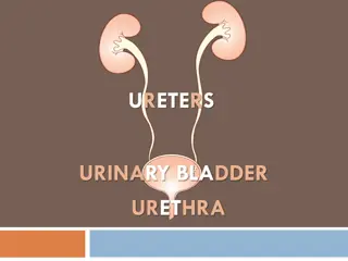
COURSE IN PELVIS In the pelvis, the ureter first runs downward, backward, and laterally along the anterior margin of the greater sciatic notch. Opposite to the ischial spine, it turns forward and medially to reach the base of the urinary bladder. Where it enters the bladder wall obliquely. Within the bladder wall, it narrows down, takes a sinuous course, and opens into the cavity of the bladder at the lateral angle of its trigone as ureteric orifice. 5
PARTS AND RELATIONS The ureter is generally divided into two parts: abdominal and pelvic. Each part is about the same length, i.e., 12.5 cm (5 inches). The abdominal part of ureter extends from the renal pelvis to the bifurcation of the common iliac artery. The pelvic part of the ureter extends from the pelvic brim (at the level of bifurcation of the common iliac artery) to the base of the urinary bladder. 6
RELATIONS OF ABDOMINAL PART Medially the right ureter is related to inferior vena cava and left ureter is related to left gonadal vein and inferior mesenteric vein. 7
RELATIONS OF PELVIC PART The pelvic part of the ureter crosses in front of all the nerves and vessels on the lateral pelvic wall except vas deferens, which crosses in front of it. Near the uterine cervix, the uterine artery lies above and in front of it, a highly important surgical relationship. 9
SITES OF ANATOMICAL NARROWINGS/CONSTRICTIONS The lumen of the ureter is not uniform throughout and presents three constrictions at the following sites. At the pelviureteric junction where the renal pelvis joins the upper end of ureter. It is the upper most constriction, found approximately 5 cm away from the hilum of kidney. At the pelvic brim where it crosses the common iliac artery. At the uretero-vesical junction (i.e., where ureter enters into the bladder). 10
In addition to above three sites of constrictions, two more sites of constrictions are described by the surgeons. At juxtaposition of the vas deferens/broad ligament. At the ureteric orifice. 11
ARTERIAL SUPPLY The ureter derives its arterial supply from the branches of all the arteries related to it. The important arteries supplying ureter from above downward are: Renal. Testicular or ovarian. Direct branches from aorta. Internal iliac. Vesical (superior and inferior). Middle rectal. Uterine. 12
VENOUS DRAINAGE The venous blood from the ureter is drained into the veins corresponding to the arteries. LYMPHATIC DRAINAGE The lymph from the ureter is drained into lateral aortic and iliac nodes. NERVE SUPPLY 1. The sympathetic supply of the ureter is derived from T12 L1 spinal segments through renal, aortic, and hypogastric plexuses. 2. The parasympathetic supply of ureter is derived from S2 S4 spinal segments through pelvic splanchnic nerves. The afferent fibres travel with both sympathetic and parasympathetic nerves. 13
Clinical Correlation Mobilization of ureter: Branches of the arteries supplying the ureter form an anastomosis in the fat and fascia around the ureter. Therefore, surgeons should bear in their mind that stripping off this fascia, while mobilizing the ureter for transplantation, will hamper the blood supply of the ureter and may cause its necrosis. Identification of ureter: Ureter is a muscular structure, and in life waves of muscular contractions produce a worm-like rhythmic movement (peristalsis) thus milking urine toward the bladder. The ureter is readily identified in life by its thick muscular wall which is seen to undergo worm-like writhing movements, especially when it is gently stroked or Squeezed. 14
Clinical Correlation Ureteric calculus is likely to lodge at one of the sites of anatomical narrowings of the ureter particularly: (a) At the pelvic ureteric junction. (b) Where it crosses the pelvic brim. (c) In the intramural part the narrowest part. Injury to ureters: According to Kenson and Hinman, the ureter may be injured at one of the following four dangerous sites: (a) Point where the ureter crosses the iliac vessels. (b) In the ovarian fossa. (c) Where the ureter is crossed by the uterine artery (most dangerous site) as damage is likely at this site during hysterectomy. (d) At the base of the bladder. 15
Thank you! 16