Veterinary Suturing Techniques and Materials Overview
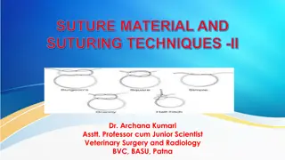
Understanding the principles of suturing in veterinary surgery is crucial for effective wound closure. This article covers different suturing techniques such as apposition, inversion, and miscellaneous sutures, along with details on needle handling, tissue penetration depth, and suture placement. Images illustrate proper needle grasping, tissue piercing angles, and specific suture patterns commonly used in veterinary practice.
Download Presentation

Please find below an Image/Link to download the presentation.
The content on the website is provided AS IS for your information and personal use only. It may not be sold, licensed, or shared on other websites without obtaining consent from the author.If you encounter any issues during the download, it is possible that the publisher has removed the file from their server.
You are allowed to download the files provided on this website for personal or commercial use, subject to the condition that they are used lawfully. All files are the property of their respective owners.
The content on the website is provided AS IS for your information and personal use only. It may not be sold, licensed, or shared on other websites without obtaining consent from the author.
E N D
Presentation Transcript
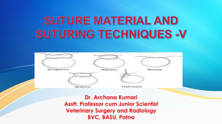
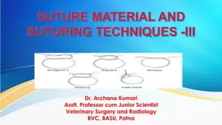
SUTURE MATERIAL AND SUTURING TECHNIQUES -V Dr. Archana Kumari Asstt. Professor cum Junior Scientist Veterinary Surgery and Radiology BVC, BASU, Patna
Principle of suturing The needle should be grasped at approximately 1/3 of the distance from the eye & 2/3 from point. The needle should be pierced the tissue perpendicular to its surface. The needle should be placed equidistant (2-3mm) from the incision line. The depth of penetration should be equal on both side of incision line. The needle always passes from The movable tissue to the fixed tissue. Thinner tissue to the thicker tissue. Deeper tissue to the superficial tissue. The tissue never be closed under tension. Each suture must be placed 3-4 mm apart from the incision line.
Suturing techniques Suture pattern are broadly classified into interrupted suture and continuous suture. Special sutures used for various purpose may also fall in one of these two categories. 1)Apposition suture 2) Inversion suture 3) Eversion suture 4) Purse-string suture 5) Relaxation suture 6) Miscellaneous suture
Apposition suture: These sutures bring about apposition of wound edges. They are commonly used for wound on skin, muscles, esophagus etc. e.g. simple interrupted, simple continuous, lock stitch, subcuticular suture and pin sutute. (i) Simple interrupted suture (ii) Simple continuous suture (iii) Continuous lock-stitch suture (iv) Sub-cuticular suture (v) Pin suture
Inversion suture In this pattern, the edges of the wound are inverted and brought together. They are used in hollow visceral organs which have a serous outer coat like intestine, uterus etc. When sutures the serous surface come in contact with each other. Inversion, sutures are effective in preventing leakage. e.g. Lembert s Czerny s, Connell and Cushing suture. i)Lembert s suture:The suture passes through serous and muscular layers but not the mucosal layer. The needle bites at right angle to the suture line. It may be continuous or interrupted pattern. ii)Czerny s suture:It is double row of lembert s suture, where one row is buried by the other row.
iii)Joberts suture: It is just like lemberts suture. Only difference is that, it penetrates all the layers. As it pierces all layers, chances of contamination is more using this suture pattern. iv)Cushing suture: This type of suture is almost similar to Lembert s suture. the difference is that suture line is parallel to the line of incision, whereas it is perpendicular in lembert s suture. It also penetrates the layers except mucous layer. v)Connell suture: The needle penetrates all the layers including mucous layer. The pattern of suturing is same as cushing.
Eversion suture The edges are everted. It is particularly used to suture cut end of the vessels. Due to eversion chances of coagulation leading to thrombi or emboli inside vessel is eliminated. Purse-string suture This suture is used to narrow the lumen of hollow organ and also to constrict the anal opening after reducing the rectal prolapse.
Relaxation suture They are usually used in large animals when there is loss of large tissues and undue tension is created on the suture line. The tension or relaxation sutures are placed relaxation at the actual line. Examples are:mattress suture, quill suture, button suture, near and far suture etc. i)Mattress of Halsted suture ii)Quill suture: These are vertical mattress suture in which pieces of quill are placed for equal distribution of tension and to prevent cutting of skin. iii)Button suture: it is also relaxation suture where buttons are used instead of quill.
Near and far suture: It is a modification of a vertical mattress suture pattern and have the advantage that is tends to prevent either inversion or eversion of the edges of the incision.
Miscellaneous suture techniques- Tendon suture (Bunnell s mayer Technique): In this technique two small atraumatic straight needles are threaded with suture material to two swaged on needles are taken. The suturing is started on proximal stump from the middle of the severed or cut end and needle is inserted teansversely through the tendon from the medial to lateral aspect and then from lateral to medial in a cross manner upto 2-3 cm proximal to the cut end and then back to the cut end to complete the first stage of suturing. The same procedure is repeated on the distal stump and the knots are tied together using the free end of the suture of the proximal and distal stumps.
Nerve suture: In the suturing of nerves the fibres are exactly oriented as far as possible and then the simple interrupted sutures are placed in the epineurium after applying the stay sutures. Staple type suture: In this technique one edge is placed over the other and sutured. Retension suture: It is used to retain gauze packing inside a wound cavity.
Parker Kerr suture: It is used to anastomose the intestine. In this method first the cut ends of the intestine are temporarily closed with the continuous Lembert s/Cusing suture by passing the loops of suture over the intestinal clamp and the sutures are kept temporarily tight. The inverted bowel ends are placed together for anastomosis and continuous Lembert s/Cusing sutures are applied to approximate the two segments. After finishing the Lembert s suture the previously placed sutures are pulled out, the continuity of double row of continuous suture are used to complete the anastomosis.
Interrupted Continuous Miscellaneous 1. Simple interrupted Simple continuous Tendon suture 2. Halsted or Mattress i. Horizontal ii. Vertical Lock stitch Nerve suture (Glover s suture) 3. Quill or Button suture Subcuticular Parker-kerr 4. Pin suture Staple stitch Lembert s 5. Relaxation or tension suture Purse string Jobert s 6. Near and far Czerny ___ 7. ___ Cushing ___ 8. ___ Connell ___
