Understanding Vestibular and Oculomotor Impairments Post-Concussion
This presentation discusses common vestibular and oculomotor impairments following a concussion, highlighting the importance of the Vestibular/Ocular Motor Screening Assessment in identifying and monitoring concussions. It covers evaluation techniques for the vestibular system, interventions for ocular and vestibular impairments, and common complaints post mild traumatic brain injury. The elements of Ocular Motor Exam are also explored in detail, along with information on ocular testing and misalignment.
Download Presentation

Please find below an Image/Link to download the presentation.
The content on the website is provided AS IS for your information and personal use only. It may not be sold, licensed, or shared on other websites without obtaining consent from the author. Download presentation by click this link. If you encounter any issues during the download, it is possible that the publisher has removed the file from their server.
E N D
Presentation Transcript
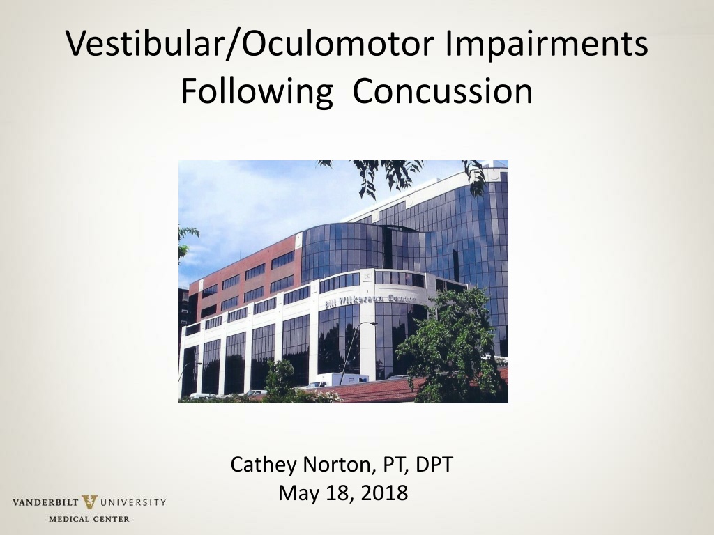
Vestibular/Oculomotor Impairments Following Concussion Cathey Norton, PT, DPT May 18, 2018
Learning Objectives Identify common vestibular and oculomotor impairments following concussion. Discuss use of the Vestibular/Ocular Motor Screening Assessment to identify concussion. Discuss evaluation of vestibular system following concussion. Present interventions for ocular and vestibular impairments.
Why Perform Oculomotor Exam? Almost 30% of concussed athletes report visual problems during the first week after the injury Vestibular and oculomotor impairment and symptoms occur in approximately 60% of athletes following SRC (Broglio 2015) 17% of athletes experience prolonged recovery lasting greater than 3 weeks (Broglio 2015) Useful in identifying concussion Useful in monitoring recovery Result of CNS decompensation pre-existing oculomotor issues may be seen (Mucha 2014)
Common Complaints following mTBI Photophobia Double vision - diplopia Blurred vision Loss of vision Visual processing problems Asthenopia eye strain Difficulty in reading Headaches Ocular pain Ventura 2014, Mucha 2014 5
Elements of OME Ocular alignment Gaze Holding Smooth Pursuits Saccades Convergence/Divergence Optokinetic nystagmus
Ocular testing Trophia always present. Can be seen in binocular vision Phoria when fusion is broken. One eye covered. Results in: Head tilt Diplopia Difficulty focusing (reading) Headaches Eye strain
Ocular misalignment Exotropia. University of Michigan Kellogg Eye Center. 26 Feb. 2013. 9
Ocular misalignment https://www.youtube.com/watch?v=cLm4o CbovsE
Testing Cover-uncover tests for trophias Alternate cover or cross cover tests for phorias. Must break fusion. Have patient wear glasses. Want best corrected vision. (Mucha 2015) Focus on a target ~16 inches in front of them.
Gaze Holding/ Fixation May indicate the presence of a central (cerebellar or brainstem) lesion Eye drifts towards center from lateral target. Corrective saccades are made to reposition target on fovea. Sedatives , anticonvulsants and recreational drugs can cause GEN http://www.dizziness-and- balance.com/practice/nystagmus/gen.htm 14
GEN https://www.youtube.com/watch?v=WJGFRTc gbOw with fixation blocked https://www.youtube.com/watch?v=rc_qfE9p 9TQ without fixation blocked
Smooth Pursuits Can cause difficulty with reading, watching moving objects TV, video games, computer, driving Tests higher level cognitive function requires attention, anticipation and working memory Ventura 2014 16
Pursuit testing Head held in neutral by examiner Object moves no more than 30 degrees/sec Move 30 degrees to each side Repeat several times response can intensify with repetition Abnormal test -inability to follow an object without saccadic eye movements
Saccades Saccades typically move at speeds between 200 and 600 degrees/sec (Hain 2014) CNS disorder - cerebellar Patient s head held on neutral position Target 20-24 from patient Fixate on a peripheral target 30 deg off midline and then a central object, examiner s nose for 10 seconds. Abnormal overshooting, need for more than 2 saccadic corrections, or gross dysconjugate eye movements (Ellis 2015)
Convergence Pulls eyes inward to focus on near target Symptoms include: Difficulty reading - comprehension Difficulty focusing from near to far Headaches Eye strain Convergence insuffiency >6 cm from tip of nose Convergence spasm increased vergence response
Convergence testing Using a single target 3 sizes above threshold near visual acuity. (Steinhafel 2015) Move target slowly toward the tip of the nose. Patient notes when target is double, not blurry. NPC- double or one eye deviates (break) Recovery- slowly pull object back until patient sees one object. 2-4 cm between break/recovery
Vestibular/Ocular-Motor Screening VOMS Developed as a clinical screen to assess and monitor vestibular and ocular motor symptoms: Items include: Smooth pursuit Horizontal and vertical saccades Convergence Accommodation Horizontal vestibular ocular reflex (VOR) Visual motion sensitivity (VMS) Post-Concussion Symptom Scale (PCSS) Mucha, et al, 2014
VOMS https://www.youtube.com/watch?v=E2uF0lcy Nps VOMS 2 is the most up to date. Addition of accommodation.
VOMS Interpretation 2 total symptoms after any VOMS item = 96% accuracy in identifying concussion NPC distance of 5 cm resulted in high rates = 84% accuracy in identifying concussion Mucha 2014
Common Oculomotor Impairments Results of VOMS study (n=85): Smooth Pursuits 33% Horizontal Saccades 42% Vertical Saccades 38% Convergence 34% VOR 58% Mucha 2014 27
http://www.moata.net/wp- content/uploads/2016/06/VOMS- scoresheet-2-cvl-edited.pdf
http://www.moata.net/wp- content/uploads/2016/06/VOMS- scoresheet-2-cvl-edited.pdf
Limitations of VOMS Validated for 9 40 year old Not a standalone test Not validated as a sideline assessment
Vestibular and Oculomotor Assessments May Increase Accuracy of Subacute Concussion Assessment SOT s ratio scores, NPC and OKS , S/S score 98.6 % accurate Optokinetic stimulation, and gaze stabilization test, S/S scores and near point convergence 94.4 % accuracy Neurocom Sensory Organization Test (SOT) Balance Error Scoring System exam 8 vestibular and oculomotor assessments. Near point of convergence Horizontal saccades Slow & fast smooth pursuits (horizontal) Optikinetic stimulation (OKS) Horizontal gaze stabilization test (GST) Head thrust (VOR test) Dynamic visual acuity (DVA) King-Devick (KD) tool McDevitt, et al, 2016
Instructions for test administration http://www.visionlink.co.nz/docs/vestibular_o cmo_screening_tool.pdf
Dizziness Dizziness is reported by 50% (Mucha 2014) to 79% (Lovell 2006) of concussed athletes 6.4x greater risk in predicting protracted (>21 days) recovery (Furman 2010) Post-concussive dizziness may arise from several sources, including benign paroxysmal positional vertigo (BPPV), post-traumatic migraines, labyrinthine concussion, perilymphatic fistula and brainstem concussion. Furman 2010 34
When to refer Abnormal alignment trophia or phoria that is not pre- existing Abnormal Saccadic , smooth pursuit or gaze holding nystagmus Visual Field cuts ex: hemianopsia Cranial nerve impairments ex: oculomotor, abducens nerve palsies Large convergence insufficiency Convergence spasm > 4 weeks or no improvement with intervention.
Vestibulo-Ocular Exam
Components of Vestibulo-Ocular Exam VOR vestibulo-ocular reflex VOR cancellation HIT head impulse test Dynamic Visual Acuity Gaze Stabilization
Vestibulo-ocular reflex (VOR) Serves vision by generating conjugate smooth eye movements. These are approximately equal and opposite in direction to head movements. As the eyes and head move in opposite directions, the ratio of eye/head velocity, the VOR gain, must approximate unity. Abnormalities cause visual blurring, oscillopsia and dizziness when the head is moving.
Spatial Arrangement of Semicircular Canals dizziness-and-balance.com 39
Head Impulse test (HIT) Can test horizontal and vertical directions 50% canal paresis is needed for a HIT to be positive. Good test for uncompensated unilateral and bilateral vestibular hypofunction Hamid 2005, 1996 40
HIT Check for adequate cervical ROM Flex neck ~ 30 degrees Patient focuses on your nose Turn head quickly ~ 10-15 degrees Test each side Unpredictable May be performed for anterior and posterior canals. + corrective (overt saccade) when turned to affected side
HIT Video https://www.youtube.com/watch?v=Wh2ojfg bC3I
Dynamic Visual Acuity Measure of gaze stability (VOR) Helps identify individuals who may have a deficit of the vestibular system http://www.nihtoolbox.org/WhatAndWhy/Se nsation/Vestibular/Pages/NIH-Toolbox- Dynamic-Visual-Acuity-Test-.aspx
Dynamic Visual Acuity Patient is seated appropriate distance from eye chart 10 ft or 20 ft. Establish static visual acuity by lowest line patient can read on eye chart Flex neck ~30 degrees Passively oscillate the head 20-30 degrees from mid line at 2 Hz (2 cycles per second) Loss of 3 or more lines indicates possible vestibular dysfunction http://www.rehabmeasures.org/Lists/Reha bMeasures/PrintView.aspx?ID=1194 44
inVision test http://www.natus.com/index.cfm?page=pr oducts_1&crid=273
GST & DVA Gaze stabilization- Quantifies the range of head movement velocities on a given axis over which a patient is able to maintain an acceptable level of visual acuity. Dynamic Visual Acuity - Quantifies the impact of Vestibular-Ocular Reflex (VOR) system impairment on a patient's ability to perceive objects accurately while moving the head at a given velocity on a given axis. http://www.natus.com/index.cfm?page=pr oducts_1&crid=273
Computerized Dynamic Platform Posturography Assess influence of the sensory system on balance Visual Vestibular Somatosensory http://www.natus.com/index.cfm?page=pr oducts_1&crid=270
Benign Paroxysmal Positional Vertigo
Benign Paroxysmal Positional Vertigo Benign paroxysmal positional vertigo (BPPV) is among the most common causes of vertigo resulting from head trauma. (Fife 2013) Recurrence rates are similar to idiopathic BPPV, but it may take more positioning maneuvers to achieve success. (Fife 2013, Liu 2012, Seong-Ki 2011) In addition, traumatic BPPV is more likely to be bilateral, occurring in 25% compared with only 2% in idiopathic BPPV. (Fife 2013)
