Understanding Stomatitis: Causes, Symptoms, and Management
Stomatitis is the inflammation of the oral mucosa, involving conditions like glossitis, palatitis, and gingivitis. It can be caused by physical, chemical, or infectious agents. Trauma, irritant drugs, and various infections contribute to this condition. Recognizing the etiology and factors contributing to stomatitis is crucial for effective management and treatment.

Download Presentation

Please find below an Image/Link to download the presentation.
The content on the website is provided AS IS for your information and personal use only. It may not be sold, licensed, or shared on other websites without obtaining consent from the author. Download presentation by click this link. If you encounter any issues during the download, it is possible that the publisher has removed the file from their server.
E N D
Presentation Transcript
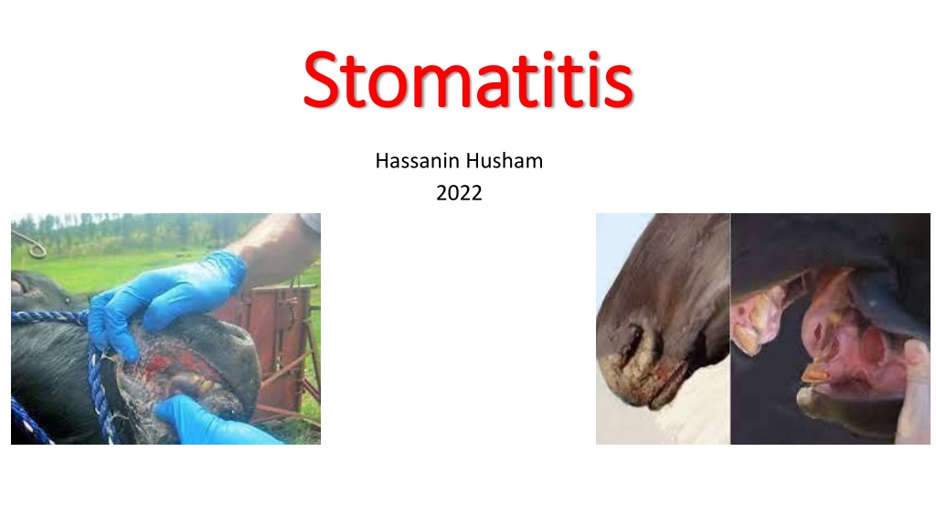
Stomatitis Stomatitis Hassanin Husham 2022
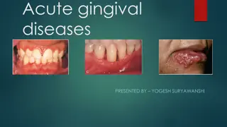
STOMATITIS STOMATITIS Stomatitis is inflammation of the oral mucosa and includes glossitis (inflammation of the tongue), palatitis (lampas; inflammation of the palate), and gingivitis (inflammation of the mucosa of the gums). Clinically it is characterized by partial or complete loss of appetite, smacking of the lips, and profuse salivation.
ETIOLOGY Stomatitis can be caused by physical, chemical, or infectious agents, with the last being the largest group of causes. The agents are listed next. 1-Physical Agents 2-Chemical Agents 3-Infectious Agents Cattle 4-Bullous Stomatitis
Physical Agents Trauma while dosing orally with a balling gun or similar instruments. Laceration of the tongue. Foreign body injury. Malocclusion of teeth. Sharp spines on plants. Eating frozen feed and drinking hot water are recorded, but seem highly improbable. Ulcers of the soft palate of horses can be caused by mechanical trauma associated with dorsal displacement of the soft palate.
Chemical Agents Irritant drugs, e.g., chloral hydrate, administered in excessive concentrations. Counterirritants applied to skin, left unprotected, and licked by the animal, including mercury and cantharides compounds. Irritant substances administered by mistake, including acids, alkalis, and phenolic compounds. Manifestation of systemic poisoning, e.g., chronic mercury poisoning and some fungi (Stachybotrys, Fusarium spp., and mushrooms) cause a combination of focal hemorrhages and necrotic ulcersor erosions. They are a common cause of confusion with vesicular or erosive disease. Lesions associated with uremia syndrome in horses.
Infectious Agents Cattle Oral necrobacillosis associated with Fusobacterium necrophorum. Actinobacillosis of the bovine tongue is not a stomatitis, Ulcerative, granulomatous lesions may occur on the gums in cases of actinomycosis. Stomatitis with vesicles occurs in vesicular stomatitis (VS). Erosive, with some secondary ulcerative, stomatitis occurs in bovine viral diarrhea (mucosal disease), bovine malignant catarrh, rinderpest. and rarely in bluetongue. Cases of infectious bovine rhinotracheitis in young calves may have similar lesions.
Proliferative lesions occur in papular stomatitis, proliferative stomatitis, and rare cases of rhinosporidiosis and papillomatosis where the oral mucosa is invaded. Oral mucosal necrosis in bovine sweating sickness. Erosive lesions in bluetongue, rinderpest, and peste de petits ruminantes. Vesicular lesions rarely in foot-and mouth disease (FMD). Granulomatous lesions caused by ecthyma are not unusual in the mouth, especially in young lambs. Cheilitis and gingivitis (inflammatory nodules of the lips and gums caused by plant awns) Lingual abscess associated with Actinobacillus spp.
Bullous Stomatitis Bullous stomatitis has been reported in the horse and can be associated with a paraneoplastic pemphigus syndrome.
PATHOGENESIS The lesions of stomatitis are produced by the causative agents being applied directly to the mucosa, or gaining entrance to it by way of minor abrasions, or by localization in the mucosa from a viremia. The clinical signs of stomatitis are caused by the inflammation or erosion of the mucosa and the signs vary in severity with the degree of inflammation.
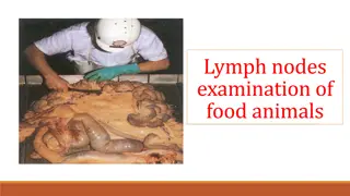
CLINICAL FINDINGS 1-There is partial or complete anorexia and slow, painful mastication. Chewing movements accompanied by salivation, animal does not swallow normally. 2- The saliva may contain pus or shreds of epithelial tissue. 3- A fetid odor is present on the breath and enlargement of local lymph nodes may also occur if bacteria invade the lesions. 4- An increased desire for water is apparent . 5- Toxemia may be present when the stomatitis is secondary to a systemic disease or where tissue necrosis occurs.
6- Traumatic lesions are usually solitary and characterized by a discontinuity in the mucous membrane often with evidence of healing and the presence of granulation tissue. 7-Catarrhal stomatitis is manifested by a diffuse inflammation of the buccal mucosa and is commonly the result of direct injury by chemical or physical agents.
8- Ulceration of the soft palate of horses with dorsal displacement of the soft palate and is characterized clinically by reduced exercise tolerance, respiratory noise during light exercise or racing, dysphagia, and coughing after exercising. . 9- Bullous stomatitis in the horse is characterized by intact or ruptured vesicles on the peripheralmargin of the tongue, the sublingual region, and the mucosa of the oral cavity and lips.
CLINICAL PATHOLOGY Material collected from lesions of stomatitis should be examined for the presence of pathogenic bacteria and fungi. Transmission experiments may be undertaken with filtrates of swabs or scrapings if the disease is thought to be caused by a viral agent.
NECROPSY FINDINGS Oral lesions are easily observed, but complete necropsy examinations should be performed on all fatally affected animals to determine whether the oral lesions are primary or are local manifestations of a systemic disease.
DIFFERENTIAL DIAGNOSIS Particularly in cattle, and to a lesser extent in sheep, the diagnosis of stomatitis is most important because of the occurrence of oral lesions in a number of highly infectious viral diseases. Careful clinical and necropsy examinations are necessary to define the type and extent of the lesions if any attempt at field diagnosis is to be made. In cattle, lymphoma of the ramus of the mandible may spread extensively through the submucosal tissues of the mouth causing marked swelling of the gums, spreading of the teeth, inability to close the mouth, and profuse salivation. There is no discontinuity or inflammation of the buccal mucosa, but gross enlargement of the cranial lymph nodes is usual. The differentiation of causes of hypersalivation must depend on a careful examination of the mouth (the causative gingivitis is often surprisingly moderate in horses) and an awareness of the volume of increased saliva output caused by toxic hyperthermia, e.g., in fescue and ergot poisonings. Poisoning by the mycotoxin slaframine also causes hypersalivation.
TREATMENT Affected animals should be isolated and fed and watered from separate utensils if an infectious agent is suspected. Nonspecific treatment application of a mild antiseptic collutory such as a 2% solution of copper sulfate, a 2% suspension of borax, or a 1% suspension of a sulfonamide in glycerin. Indolent ulcers require more vigorous treatment and respond well to curettage or cauterization with a silver nitrate stick or tincture of iodine. In stomatitis caused by trauma, the teeth might need attention. In all cases, soft, appetizing food should be offered. If the disease is infectious, care should be exercised to ensure that it is not transmitted by the hands or dosing implements.