Understanding Protein Synthesis and Translation Process
Proteins play a crucial role in various body structures and functions. The process of translation, as per the central dogma, involves converting genetic information from DNA to functional proteins through transcription and translation. This process includes initiation, elongation, and termination steps with the participation of different types of RNAs. The initiation step involves tRNA charged with methionine binding to the mRNA's AUG codon, forming the initiation complex on the ribosome. Subsequent steps include the introduction of charged tRNA to the P site and A site, determining the incorporation of amino acids based on triplet codons. Understanding these processes is essential for grasping how proteins are synthesized in the body.
Download Presentation

Please find below an Image/Link to download the presentation.
The content on the website is provided AS IS for your information and personal use only. It may not be sold, licensed, or shared on other websites without obtaining consent from the author. Download presentation by click this link. If you encounter any issues during the download, it is possible that the publisher has removed the file from their server.
E N D
Presentation Transcript
Lecture 13 :Protein synthesis TRANSLATION
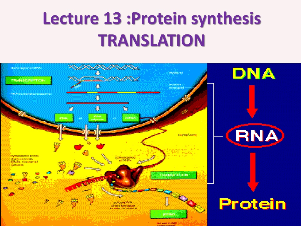
According to central dogma , the genetic information flow from DNA the RNA via transcription process then it will translate later to functional protein.
TRANSLATION In this process the three types of RNA are involved. Each one of them plays an important role to complete protein synthesis . The translation also involves 1-Initiation 2- Elongation 3- Termination step
Step 1- 2- tRNA which will come charged (bind to A.A) and the first A.A is methionine (Met) because the first codon is AUG become as formyl methionine). The AUG first codon in mRNA will face the triplet codon in anti 3- small 30S ribosomal sub units thus the m RNA will bind to 16s rRNA via its ribosomal binding site . one: Initiation , it requires cell) mRNA (processed in Eukaryotic (usually codon arm in tRNA.
The initiation step requires initiation factors then the large subunit bind the complex forming 70s initiation complex. The large subunit contain P site (Peptidyl sit ) and A site (Amino -acyl site ).
The first occupied site by the charged tRNA is P site followed by introducing A site and the triplet codon will determine the next incorporated A,A
The 4 nitrogen base will form 64 possibilities and we have 20 A.A thus 2-3 triplet codon will code to the same A,A. beside we have one start codon (AUG) and 3 stop codon (UAA, UAG ,UAG) that they will not translated to any A.A.
2- Elongation step :here the peptide chain will elongate with the help of elongation factors. The A.A in the p site will bind with the A.A in A site via peptide bond with the add of peptidyl transferase. tRNA in p site now is empty, converts to uncharged and leave the ribosome from Exit site. Then the tRNA translocats from A to P site with 2 A.A . Now the A site is empty and the complex will move for another triplet codon
The figure shows ribosome movement (from 53) as each 3 nitrogen base form one triplet codon and will translate to one A.A. the growing peptide chain will continue move between P and A site along them RNA untill reaching the stop codon.
Termination step: when the ribosome reached stop codon (UAG, UAA, UGA) and there is no anticodon in tRNA so A site will remain empty causes releasing of free polypeptide and dissociation of the 2 ribosomal subunits. Note: mRNA will degraded once it translated thus its concretion is low while m RNA in Eukaryotic cell more stable because it contains cap structure at 5 end and poly A tail in 3 end .
Basic structure of protein: the structural unit of protein is amino acid (A.A).The main structure of A.A is 1- central C atom called - carbon. . 2-Amino group gives the positive charge 3- carboxylic acid group gives the negative charge 4- R side chain which differ from one amino acid to another, the simplest R side group is H only to form Glycin
The bond which bind the two adjacent A.A is called peptide bond with releasing of water molecules .
Usually the protein chain start with amino group (NH3) and end with Carboxyl group (COOH). Binding of 2-10 a.a gives a rise to oligopeptide while binding more than that will form the polypeptide chain. This linear chain represent 1- the primary structure to the protein.
According to structure and configuration ,proteins can be divided to 4 types
2- The secondary structure formed due to folding of the primary structure via hydrogen bonds. Two types are well studied as secondary structur 1- helix pleated sheet 2-
3- Tertiary structure is more complicated and we could find helix and pleated sheet in the same structure ,other types of bonds exist here like disulfide bond between cystine A.A ,ionic bond ,hydrophobic bond
4- Quaternary structure: more complicated result from folding the other structures and from more than subunits
