Understanding Mitotic Index in Practical Biology
Explore the practical technique of determining the mitotic index, a key aspect of cell division, through a step-by-step process involving garlic root tip preparation and microscopic observation. Discover real-world applications and enhance your skills in risk assessment and tissue analysis. Uncover the fascinating world of meristematic tissue and chromosomal visualization in biological studies.
Download Presentation

Please find below an Image/Link to download the presentation.
The content on the website is provided AS IS for your information and personal use only. It may not be sold, licensed, or shared on other websites without obtaining consent from the author. Download presentation by click this link. If you encounter any issues during the download, it is possible that the publisher has removed the file from their server.
E N D
Presentation Transcript
Required Practical 2: Mitotic Index
What is a real-world application of this practical technique?
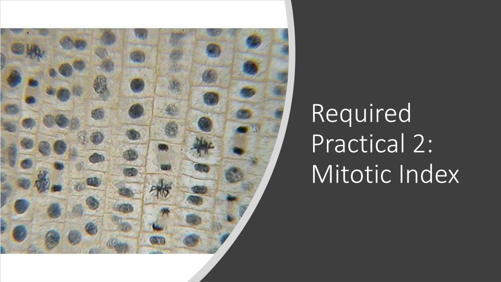
Key Term: Meristem Meristematic Tissue How do we get to the point where we can see the garlic chromosomes under an optical microscope?
New Skill: Writing a Risk Assessment (C3a)
Method Fill 25ml beaker with 10ml Hydrochloric acid, place in water bath and leave for 15mins to allow acid to warm up to 55oC. Place garlic clove in acid, so that the root tips are submerged in acid. Leave roots in acid for 5 mins. Carefully remove garlic from acid and rinse in beaker of water. Place slide on white tile. Cut off the root tip (approx. 1mm) on to slide. Cover cells with a drop of toludiene blue, and macerate with mounted needle to separate cells. Continue to macerate until the tissue is well broken down and the cells are stained dark blue. Add a cover slip and with gentle finger pressure spread the material, blotting any stain that leaks out using filter paper. Look carefully at slides for on-going mitosis under a microscope. Chromosomes should stain dark blue. Repeat for several fields of view. Record your data in a suitable table. Calculate the mitotic index.
