Understanding Hemoglobin: Synthesis, Functions, and Influence on Anemia
Hemoglobin is a vital protein in red blood cells responsible for oxygen transport. Its synthesis, influenced by factors like erythropoietin, vitamins, and trace metals, plays a crucial role in maintaining proper oxygen levels in the body. An insight into hemoglobin's structure, synthesis, and impact on conditions like anemia can help in comprehending its significance in maintaining overall health.
Download Presentation

Please find below an Image/Link to download the presentation.
The content on the website is provided AS IS for your information and personal use only. It may not be sold, licensed, or shared on other websites without obtaining consent from the author. Download presentation by click this link. If you encounter any issues during the download, it is possible that the publisher has removed the file from their server.
E N D
Presentation Transcript
Lab# 5 BCH 471 Hemoglobin and anemia
Objectives: 1- Quantitative determination of hemoglobin in a blood sample. 2- Quantitative determination of glucose 6-phosphate dehydrogenase (G6PD) activity in erythrocytes (hemolysate). 3- Qualitative determination of hemoglobin S (HbS) in blood using a phosphate solubility method.
Hemoglobin: Hemoglobin the RBCs (erythrocytes) that transport oxygen from the lungs to the rest of the body and carbon dioxide back to the lungs. (Hb),is the iron-containing oxygen-transport metalloprotein in The protein part: 4 subunits of globin protein. The non-protein part: heme group (iron + Protoporphyrin). Hb is made up of protein and non-protein parts.
Hemoglobin Synthesis The circulation blood of normal adult contain about 750 g of Hb and of this about 7 8 g are degraded daily. The globin part of Hb can be reutilized only after catabolism into its constituent amino acid. The free heam is broken down into bile pigment which is excreted. Iron alone is reutilized in the synthesis of Hb. The rate of Hb synthesis (Rate of RBC formation) depends on: The amount of oxygen reaching the blood. Capacity of the blood to carry oxygen ,which in turn depend on the amount of circulating hemoglobin
Therefore, hemoglobin synthesis is stimulated by anoxia, whether due to oxygen deficiency or due to anemia. There is a strong evidence that the marrow response to the stimulus of hypoxia is dependent upon erythropoietin. Erythropoietin is a glycoprotein hormoneformed in kidney in response to decrease oxygen carrying capacity (hypoxia or anoxia), in order to stimulate the erythropoiesis
The role of some factor affecting on the native of haemoglobin: 1) Vitamins and cofactor: Biotin (B7), pantothenic acid (B5), folic acid (B9), coenzyme A and pyrodixal phosphate are essential for haem synthesis . 2) Trace metals : Only copper and cobalt are known to play a role . (Copper is playing a role in the absorption of iron while Cobalt is essential constituent of vitamine B12 (Cobalamin) ) 3) Glucose -6-phsphatase dehydrogenase (G6PD)
Estimation of blood haemoglobin: Principle: The ferrouse (Iron II) in each haem in RBC is oxidized by ferricyanide to Fe(III)- methaemoglobin . A cynide group (CN-) is then attached to the iron atom (because it is positively charge) by reaction with KCN to give the brown cyanomethamoglobin (stable) which can be estimated quantitatively Normal Hb conc.: for men: 14 - 18 g/dl, for women : 12 - 16 g\dl Level of Hb is associated with polycythemia and dehydration. Level of Hb is associated with anemia.
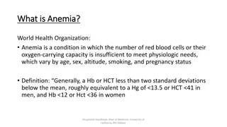
Anemia : It is in general decrease in the amount of RBC or the normal amount of Hb in blood. It can also be defined as a lowered ability of the blood to carry oxygen. There are many types of anemia: 1- Anemia caused by blood loss. 2- Anemia caused by decreased or faulty red blood cell production. (Sickle cell anemia Iron-deficiency anemia and Vitamin-deficiency anemia) 3- Anemia caused by destruction of red blood cells.(hemolytic anemia).
Quantitative Determination of G6PD Deficiency in Hemolysed RBC sample
Glucose-6-phosphate dehydrogenase, deficiency is a genetic disorder that occurs almost exclusively in males. This condition mainly affects red blood cells, which carry oxygen from the lungs to tissues throughout the body. In affected individuals, a defect in an enzyme called glucose-6-phosphate dehydrogenase causes red blood cells to break down prematurely. This destruction of red blood cells is called hemolysis. The most common medical problem associated with glucose-6-phosphate dehydrogenase deficiency is hemolytic anemia, which occurs when red blood cells are destroyed faster than the body can replace them. This type of anemia leads to paleness, yellowing of the skin and whites of the eyes (jaundice), dark urine, fatigue, shortness of breath, and a rapid heart rate.
Importance of G6PD G6PD is the enzyme responsible for the initial deviation of glucose into pentose phosphate pathway to form 6-phosphogluconate. This pathway provides NADPH2 in the erythrocyte for the conversion of oxidized to reduced glutathione and for other reactions such as reduction of methemoglobin. Deficiency of G6PD: 1- The enzyme deficiency may cause haemolysis, but chiefly occurs after sensitization of the erythrocyte by a wide variety of agents e.g. primaquine, broad beans (favism) or in infections. 2- The cells accumulate methemoglobin and are deficient in reduced glutathione which is necessary for cell integrity. (Haemolysis will cause, dark urine and jaundice). 3- In homozygote, enzyme activity is reduced to less than 15% of normal. 4- The deficiency of G6PD may also produce neonatal jaundice.
GSH is necessary for cell integrity by neutralizing free radicals that cause oxidative damage.
Principle: Erythrocytes are lysed (by saponin) and their content is released Glucose + NADP+ G6PD 6-Phosphogluconate + NADPH + H+ The rate of formation of NADPH is a measure of the G6PD activity and it can be followed by means of the increase in the Absorbance at 340 nm. Note: A red cell hemolysate is used to assay for deficiency of the enzyme, while serum is used for evaluation of enzyme elevations.
Qualitative determination of hemoglobin S (HbS) in blood. (Sickle cell test)
Hemoglobin S (Hgb S), is an abnormal type of hemoglobin that you can inherit from parents. Sickle cell test , is a simple and rapid method for detection of the presence of hemoglobin-S in blood. In normal adults, 95% or more of the hemoglobin is present as hemoglobin A (HbA). -Hemoglobin-S, can be inherited in the homozygous state (S/S) produce sickle cell anemia , or in heterozygous ,also called sickle cell trait (A/S) ), usually don t exhibit symptoms of the sickle cell anemia disease (unless under extreme hypoxia). -A point mutation in the Hb gene is responsible for the sickling of RBCs seen in sickle cell anemia .The abnormality is due to Substitution of non polar valine for a charged Glutamic acid in position 6 in the chain .
Individuals with Hb-S will be at high risk when exposed to conditions of low oxygen tension such as surgery, high altitude or athletics which may results in serious and fatal clinical complications. In order to avoid or minimize these clinical complications, it is important to screen the individuals for the presence of Hb-S. Hemoglobin A Hemoglobin A2 Hemoglobin F
Principle: Erythrocytes are lysed (by saponin) and the released hemoglobin is reduced (by dithionite) in phosphate buffer. Reduced HbS is characterized by its very low solubility , So that in the presence of HbS, the solution become turbid and the lines behind the test tube will not be visible while, if no HbS was present the clear solution will permit the lines to be seen through the test tubes.
Results: _ +
