Understanding Enzyme Inhibition and Its Kinetics
Learn about enzyme inhibitors and their impact on catalysis, the different types of reversible enzyme inhibition, including competitive and uncompetitive inhibition, and how they affect enzyme kinetics. Discover how these inhibitors work and their implications in drug design and biochemical studies.
Download Presentation

Please find below an Image/Link to download the presentation.
The content on the website is provided AS IS for your information and personal use only. It may not be sold, licensed, or shared on other websites without obtaining consent from the author. If you encounter any issues during the download, it is possible that the publisher has removed the file from their server.
You are allowed to download the files provided on this website for personal or commercial use, subject to the condition that they are used lawfully. All files are the property of their respective owners.
The content on the website is provided AS IS for your information and personal use only. It may not be sold, licensed, or shared on other websites without obtaining consent from the author.
E N D
Presentation Transcript
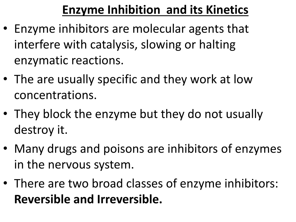
Enzyme Inhibition and its Kinetics Enzyme inhibitors are molecular agents that interfere with catalysis, slowing or halting enzymatic reactions. The are usually specific and they work at low concentrations. They block the enzyme but they do not usually destroy it. Many drugs and poisons are inhibitors of enzymes in the nervous system. There are two broad classes of enzyme inhibitors: Reversible and Irreversible.
Reversible inhibition : The inhibitor binds non-covalently with enzyme and the enzyme inhibition can be reversed if the inhibitor is removed. The reversible inhibition is further sub-divided into : l. Competitive inhibition ll. Non-competitive inhibition III. Un -competitive inhibition
1. Competitive Inhibition : These compete with the substrate molecules for the active site. Inhibitor (I) occupies the active site by preventing the binding of the substrate to the enzyme. Many competitive inhibitors are compounds that resemble the substrate and combine with the enzyme to form an EI complex, but without leading to catalysis. The inhibitor s action is proportional to its concentration.
Competitive inhibition can be analyzed quantitatively by steady-state kinetics. In the presence of a competitive inhibitor, the Michaelis-Menten equation becomes : Vo = Vmax [s] / Km + [S] where = 1+ [I]/ KI KI = [E] [I] / [EI] The experimentally determined variable Km, the Km observed in the presence of the inhibitor, is often called the apparent Km.
2. Uncompetitive inhibition : Inhibitor binds at a site distinct from the substrate active site and, unlike a competitive inhibitor, binds only to the ES complex. In the presence of an uncompetitive inhibitor, the Michaelis-Menten equation is altered to : Vo = Vmax [s] / Km + [S] where = 1+ [I]/ K I K I = [ES] [I] / [ESI]
As described by Equation, at high concentrations of substrate, Vo approaches Vmax/ . Thus, an uncompetitive inhibitor lowers the measured Vmax. Apparent Km also decreases, because the [S] required to reach one-half Vmax decreases by the factor. Both Km & Vmax decreases.
3. Non competitive Inhibition : (Mixed Inhibition) Inhibitor binds at a site distinct from the substrate active site, but it binds to either E or ES. The rate equation describing mixed inhibition: Vo = Vmax [s] / Km + [S] A mixed inhibitor usually affects both Km and Vmax.
Km remain same while Vmax changed, Vmax decreases. E.g. Heavy metals can noncompetitively inhibits the enzyme by binding with sulfhydryl group.
Irreversible Inhibition : The irreversible inhibitors are those that bind covalently with or destroy a functional group on an enzyme that is essential for the enzyme s activity, or those that form a particularly stable noncovalent association. Formation of a covalent link between an irreversible inhibitor and an enzyme is common.
Irreversible inhibitors are another useful tool for studying reaction mechanisms. Amino acids with key catalytic functions in the active site can be identified by determining which residue is covalently linked to an Inhibitor. e.g. (1) DiIsopropyl fluorophosphates reacts irreversibly with serine proteases such as chymotrypsin. (2) Idoacetate reacts with essential sulfhydryl group of Triose phosphate Isomerase.
Regulatory Enzymes : Regulatory enzymes exhibit increased or decreased catalytic activity in response to certain signals. In most multienzyme systems, the first enzyme of the sequence is a regulatory enzyme. This is an excellent place to regulate a pathway. The activities of regulatory enzymes are modulated in a variety of ways.
Allosteric enzymes function through reversible, noncovalent binding of regulatory compounds called allosteric modulators or allosteric effectors, which are generally small metabolites or cofactors. Other enzymes are regulated by reversible covalent modification. Both classes of regulatory enzymes tend to be multisubunit proteins. In some cases the regulatory site(s) and the active site are on separate subunits.
Metabolic systems have at least two other mechanisms of enzyme regulation. Some enzymes are stimulated or inhibited when they are bound by separate regulatory proteins. Others are activated when peptide segments are removed by proteolytic cleavage. Unlike effector-mediated regulation, regulation by proteolytic cleavage is irreversible.
Allosteric Enzymes : Allosteric enzymes are those having other shapes or conformations induced by the binding of modulators. The modulators for allosteric enzymes may be inhibitory or stimulatory. Often the modulator is the substrate itself. Regulatory enzymes for which substrate and modulator are identical are called homotropic. When the modulator is a molecule other than the substrate, the enzyme is said to be heterotropic
The properties of Allosteric enzymes : The properties are significantly different from those of simple nonregulatory enzymes. Some of the differences are structural. In addition to active sites, allosteric enzymes generally have one or more regulatory, or allosteric, sites for binding the modulator. each regulatory site is specific for its modulator.
Enzymes with several modulators generally have different specific binding sites for each. In homotropic enzymes, the active site and regulatory site are the same. Allosteric enzymes are generally larger and more complex than nonallosteric enzymes. Most have two or more subunits
In some multienzyme systems, the regulatory enzyme is specifically inhibited by the end product of the pathway whenever the concentration of the end product exceeds the cell s requirements. This type of regulation is called feedback inhibition. One of the first known examples of allosteric feedback inhibition was the bacterial enzyme system that catalyzes the conversion of L- threonine to L-isoleucine in five steps.
the first enzyme threonine dehydratase, is inhibited by isoleucine the product of the last reaction of the series. This is an example of heterotropic allosteric inhibition.
Isoleucine binds not to the active site but to another specific site on the enzyme molecule, the regulatory site. This binding is noncovalent and readily reversible. If the isoleucine concentration decreases, the rate of threonine dehydration increases.
The Kinetic Properties of Allosteric Enzymes : Allosteric enzymes show relationships between Vo and [S] that differ from Michaelis-Menten kinetics. They do exhibit saturation with the substrate when [S] is sufficiently high. For some allosteric enzymes, plots of Vo /[S] produce a sigmoid saturation curve, rather than the hyperbolic curve
On the sigmoid saturation curve we can find a value of [S] at which Vo is half- maximal, but we cannot refer to it with the designation Km, because the enzyme does not follow the hyperbolic Michaelis- Menten relationship. Sigmoid kinetic behaviour generally reflects cooperative interactions between protein subunits.
i.e changes in the structure of one subunit are translated into structural changes in adjacent subunits is due to noncovalent interactions at the interface between subunits. Homotropic allosteric enzymes generally are multisubunit Proteins. The same binding site on each subunit functions as both the active site and the regulatory site.
Most commonly, the substrate acts as a positive modulator (an activator), because the subunits act cooperatively: the binding of one molecule of substrate to one binding site alters the enzyme s conformation and enhances the binding of subsequent substrate molecules. This accounts for the sigmoid rather than hyperbolic change in Vo with increasing [S].
One characteristic of sigmoid kinetics is that small changes in the concentration of a modulator can be associated with large changes in activity. Heterotropic allosteric enzymes are those whose modulators are metabolites other than the normal substrate. it is difficult to generalize about the shape of the substrate-saturation curve. A negative modulator (an inhibitor) may produce a more sigmoid substrate-saturation curve.
Heterotropic allosteric enzymes therefore show different kinds of responses in their substrate-activity curves, because some have inhibitory modulators, some have activating modulators, and some have both. Sigmoid kinetic behaviour is explained by the concerted and sequential models for subunit interactions.
Concerted model : The first model was proposed by Jacques Monod,Jeffries Wyman, and Jean-Pierre Changeux in 1965. The concerted model assumes that the subunits of a cooperatively binding protein are functionally identical, that each subunit can exist in (at least) two conformations, and that all subunits undergo the transition from one conformation to the other simultaneously. In this model, no protein has individual subunits in different conformations. The two conformations are in equilibrium. The ligand can bind to either conformation, but binds each with different affinity. Successive binding of ligand molecules to the low-affinity conformation (which is more stable in the absence of ligand) makes a transition to the high-affinity conformation more likely.
sequentialmodel concerted model
Sequential model : . Proposed in 1966 by Daniel Koshland and colleagues. Ligand binding can induce a change of conformation in an individual subunit. A conformational change in one subunit makes a similar change in an adjacent subunit, as well as the binding of a second ligand molecule, more likely. There are more potential intermediate states in this model than in the concerted model. The concerted model may be viewed as the all-or-none limiting case of the sequential model.
Regulatory Enzymes Undergo Reversible Covalent Modification : Another important class of regulatory enzymes, activity is modulated by covalent modification of the enzyme molecule. Modifying groups include phosphoryl, adenylyl, uridylyl, methyl, and adenosine diphosphate ribosyl groups. These groups are generally linked to and removed from the regulatory enzyme by separate enzymes. An enzyme regulated by methylation is the methyl- accepting chemotaxis protein of bacteria. ADP-ribosylation is a type of modification occurs for the bacterial enzyme dinitrogenase reductase, resulting in regulation of the important process of biological nitrogen fixation.
Phosphorylation : is the most common type of regulatory modification; one- third to one-half of all proteins in a eukaryotic cell are phosphorylated. The attachment of phosphoryl groups to specific amino acid residues of a protein is catalyzed by protein kinases; removal of phosphoryl groups is catalyzed by protein phosphatases. phosphorylation can have dramatic effects on protein conformation and thus on substrate binding and catalysis. An important example of regulation by phosphorylation is seen in glycogen phosphorylase (Mr 94,500) of muscle and liver. Catalyzes the reaction :
Glycogen phosphorylase occurs in two forms: the more active phosphorylase a and the less active phosphorylase b . Phosphorylase a has two subunits, each with a specific Ser residue that is phosphorylated at its hydroxyl group. These serine phosphate residues are required for maximal activity of the enzyme.
The phosphoryl groups can be hydrolytically removed by a separate enzyme called phosphorylase phosphatase: In this reaction, phosphorylase a is converted to phosphorylase b by the cleavage of two serine phosphate covalent bonds, one on each subunit of glycogen phosphorylase.
Phosphorylase b can in turn be reactivatedcovalently transformed back into active phosphorylase a by another enzyme, phosphorylase kinase, which catalyzes the transfer of phosphoryl groups from ATP to the hydroxyl groups of the two specific Ser residues in phosphorylase b. The a and b forms differin their secondary, tertiary, and quaternary structures. The active site undergoes changes in structure and, consequently, changes in catalytic activity as the two forms are interconverted. The regulation of glycogen phosphorylase by Phosphorylation illustrates the effects on both structure and catalytic activity of adding a phosphoryl group.
Zymogen: An inactive precursor of some enzyme is called a zymogen, is cleaved to form the active enzyme. Many proteolytic enzymes (proteases) such as Chymotrypsin and trypsin are initially synthesized as chymotrypsinogen and trypsinogen. Specific cleavage causes conformational changes that expose the enzyme active site. This type of activation is irreversible. Proteases are not the only proteins activated by proteolysis. In other cases, the precursors are called not zymogens but, more generally, proproteins or proenzymes . E.g. protein collagen is initially synthesized as the soluble precursor procollagen. Fibrin, the protein of blood clots, is produced by proteolysis of fibrinogen, its inactive proprotein.
Isoenzymes : The same reaction catalyzed by two or more different molecular forms of an enzyme. These multiple forms, called isozymes or isoenzymes, may occur in the same species, in the same tissue, or even in the same cell. The different forms of the enzyme generally differ in kinetic or regulatory properties, in the cofactor they use. Isozymes may have similar, but not identical, amino acid sequences, and in many cases they clearly share a common evolutionary origin. First enzymes found to have isozymes was Lactate dehydrogenase (LDH) . exists as five different isozymes separable by electrophoresis. All LDH isozymes contain four polypeptide chains (each of Mr 33,500), each type containing a two kinds of polypeptides. The M (for muscle) chain and the H (for heart) chain, encoded by two different genes. In skeletal muscle isozyme contains four M chains, and in heart the predominant isozyme contains four H chains.

