Understanding Diffusion MRI in Radiology
Explore the basics of Diffusion MRI, including principles of DWI, states of restricted diffusion, and diagnostic applications in cerebral infarction. Learn about the ADC Map and interpretation of images for different brain conditions, such as neonatal brain abnormalities. Gain insights into the appearance of lesions in DW MRI and the significance of diffusion characteristics in various pathologies.

Download Presentation

Please find below an Image/Link to download the presentation.
The content on the website is provided AS IS for your information and personal use only. It may not be sold, licensed, or shared on other websites without obtaining consent from the author. If you encounter any issues during the download, it is possible that the publisher has removed the file from their server.
You are allowed to download the files provided on this website for personal or commercial use, subject to the condition that they are used lawfully. All files are the property of their respective owners.
The content on the website is provided AS IS for your information and personal use only. It may not be sold, licensed, or shared on other websites without obtaining consent from the author.
E N D
Presentation Transcript
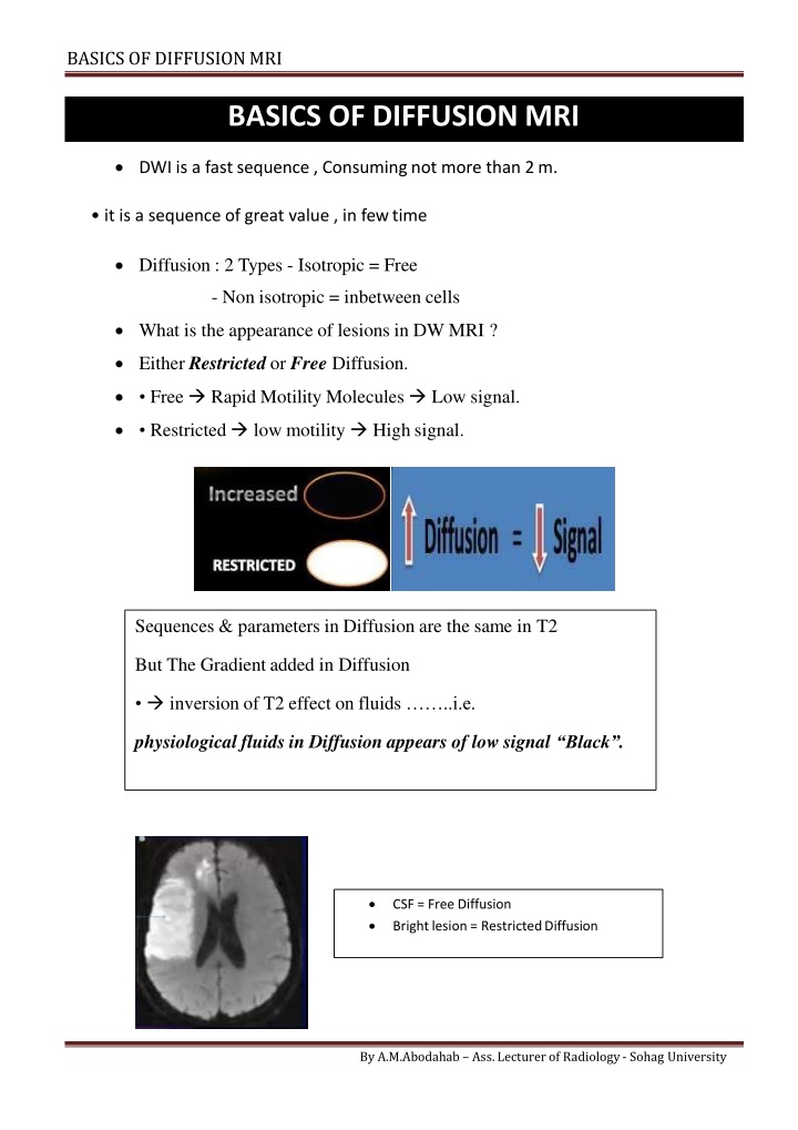
BASICS OF DIFFUSIONMRI BASICS OF DIFFUSION MRI DWI is a fast sequence , Consuming not more than 2m. it is a sequence of great value , in fewtime Diffusion : 2 Types - Isotropic = Free - Non isotropic = inbetween cells What is the appearance of lesions in DW MRI ? Either Restricted or Free Diffusion. Free Rapid Motility Molecules Low signal. Restricted low motility High signal. Sequences & parameters in Diffusion are the same in T2 But The Gradient added in Diffusion inversion of T2 effect on fluids ..i.e. physiological fluids in Diffusion appears of low signal Black . CSF = Free Diffusion Bright lesion = RestrictedDiffusion By A.M.Abodahab Ass. Lecturer of Radiology - Sohag University
BASICS OF DIFFUSIONMRI * What are the pathological principles of DWI ? Fluid Molecules diffuse in between Cells Thus, . increase in Cells Size - Number - Viscosity of intercellular Fluid DECREASE Restrict Diffusion What are the main 3 states of Restricted Diffusion ? Increase Cell Size as in Cerebral Infarction Increase Cell Number as in Tumors Increase Viscosity of intercellular Fluid As in Brain Abscess By A.M.Abodahab Ass. Lecturer of Radiology - Sohag University
BASICS OF DIFFUSIONMRI CerebralInfarction In cerebral infarction Cut off blood supply disturbance of salts exchange Na in ward Cells swollen i.e. cytotoxic edema Swelling of the cells = increase its size Diffusion Restriction .DWI is the Fastest method to diagnose Cerebral Infarction 1st 3 Hours are the golden time for Thrombolytictherapy DWI can diagnose infarction in hyper acute stage This early state not diagnosed by CT or may Not by MRI BUT DWI can diagnose in WITH IN MINUTES of occurrence!!! Infarction Stage Hyper-acute Acute Sub-acute Chronic Duration < 6 H 6 h : 3 Days 3 days : 3 weeks >3 weeks By A.M.Abodahab Ass. Lecturer of Radiology - Sohag University
BASICS OF DIFFUSIONMRI ADCMAP ADC = Apparent Diffusion Coefficient it represent the rate of an anisotropic diffusion. What is b Factor ? = Degree of diffusion mm2/sec it depend on : Gradientamplitude Time intervalsin-between .Thus , More b Factor . More Chance of Diffusion ADC Map is a computerizedimage. Obtained by taking multiple Diffusion images on different b Factors. This series of images make the rate of diffusion of different molecules can be calculated in numbers. The ADC Map image is inverted in colors . i.e. restricted is black & Free is White By A.M.Abodahab Ass. Lecturer of Radiology - Sohag University
BASICS OF DIFFUSIONMRI Normal neonatal brain. normal to have low DW signal intensities in the frontal deep white matter (arrows.( The appearance of the pediatric brain on DW images varies withage b - ADC values of the corresponding areas are high in neonatalbrain, = To interpret ADC Image: Look at : Diffusion Image & ADCMap Area of Restricted Diffusion: -High signal in DWI & Low in ADC ..& Vice versa Thus As regarding DWI . Restricted Diffusion & lesions of very high signal in T2 appears Bright How to Differentiate? Area of Restriction Bright in DWI Dark in ADC Very Hi T2 lesion Bright in DWI Bright in ADC By A.M.Abodahab Ass. Lecturer of Radiology - Sohag University
BASICS OF DIFFUSIONMRI Value of Diffusion in ADC Map is represented by ADCValue. ADC Value & Tumors Malignant Vs Benign Low grade Vs HighGrade Higher Cell number More Restricted Diffusion Less ADC Value ADC Value & Tumor Cellularity? Tumors of High Cellularity: Lymphoma ADC Value 0.51 : 0.71 High grade Glioma 0.58 :0.88 Metastasis Medulloblastoma Tumors of Low Cellularity : Low grade Glioma .. ADC Value >1.05 By A.M.Abodahab Ass. Lecturer of Radiology - Sohag University
BASICS OF DIFFUSIONMRI All Malignancy has High Cellularity ? ? UsuallyYes But Few Malignancies has Low Cellularity . Best Example CHORDOMA Chordoma has low celularity High ADCvalue Epidermoid Cysts Benign & non-neoplastic , Congenital or Acquiredcysts. Contain dense fluid + epidermal elements restricteddiffusion. DWI differentiate it from other cysts, especially : arachnoid cyst, do not show any restricted diffusion. "CSFLIKE" By A.M.Abodahab Ass. Lecturer of Radiology - Sohag University
BASICS OF DIFFUSIONMRI By A.M.Abodahab Ass. Lecturer of Radiology - Sohag University
BASICS OF DIFFUSIONMRI Meningiomas Extra-axial DuralNeoplasm Homogenous intense enhancing +/- Dural tail. Their typical appear easier to be diagnosed on routine MRimages. Subtypes: * Typical, * Atypical *Malignant A typical and malignant meningiomas show much more prominent restricted diffusion. Meningioma. Post-contrast T1-weighted image (A) shows intense enhancement of .)the tumor(arrow On ADC map, the mass is dark, due to the restricted diffusion. Pathology:Atipic .meningioma, WHO grade2 By A.M.Abodahab Ass. Lecturer of Radiology - Sohag University
BASICS OF DIFFUSIONMRI Chordomas Chordomas Vs chondro-sarcomas Rare Primary bone tumors it Involves: skull base, especially the clivus. sacrococcigeal region. DWI: Especially poorly differentiated chordomas more restricted diffusion than chondro- sarcomas By A.M.Abodahab Ass. Lecturer of Radiology - Sohag University
BASICS OF DIFFUSIONMRI Lymphomas Lymphoma Vs Glial Tumors. Site: periventricular/sub ependymal. Intense Homogenous enhancement. Their diffusion is more restricted compared to glial tumors Also, they show lower perfusion than glial tumors. Abscess . Sometimes can be mistaken as necrotic tumors on imaging, Both show peripheral contrast enhancement. Clinical presentation is important for the differential diagnosis. On MRI, liquid content of the abscesss how markedly restricted diffusion , extremely helpful for the diagnosis . By A.M.Abodahab Ass. Lecturer of Radiology - Sohag University
BASICS OF DIFFUSIONMRI Choroid Plexus Cyst Diffusion-weighted MRI (DWI) has been applied to extracranial sites since the 1990s. Several investigators have reported that 3-T DWI can improve : the diagnostic accuracy of tumor detection, Assessment of HepaticTumors staging, Assessment of body lymphoma targetedbiopsy, Evaluation of ProstateCancer posttreatment follow-up, Assessment of therapeuticresponse By A.M.Abodahab Ass. Lecturer of Radiology - Sohag University
BASICS OF DIFFUSIONMRI SUMMARY DWI is a fast sequence * Gradient its magic ink DWI is the best to assess hyper acuteinfarction Restricted diffusion = high signal in DWI / Low in ADC vice versa Increase in cells size/Infarction- Numbers / Tumors "Fluid Viscosity / Cyto-toxic edema" ADC Map give a numerical values as in CT Hu. Highest ADC value of freeDiffusion is 3 ADC value of : high grade Tumors < 1 , Low grade >1 DWI is an important part of MR imaging for the evaluation of brain masses. DWI can not be used alone. Data obtained from routine T1, T2 and FLAIR sequences as well as post contrast images should be evaluated altogether. Perfusion imaging evaluate vascularity of masses. By A.M.Abodahab Ass. Lecturer of Radiology - Sohag University


