Understanding Biliary Tract Anatomy and Interventional Procedures
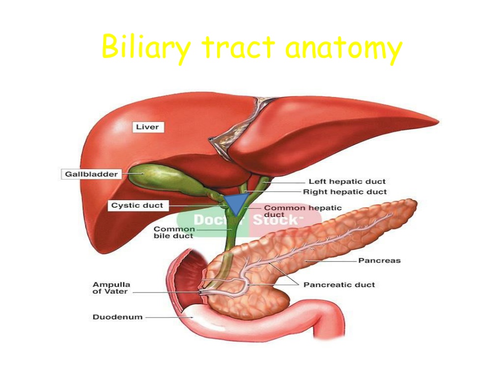
Explore the anatomy of the biliary tract, including pathways of cholesterol metabolism in the liver and the composition of bile. Learn about conditions such as gallstones, polyps, and common bile duct abnormalities, as well as diagnostic procedures like ERCP. Discover indications for biliary stenting and gain insights into managing biliary obstructions and related complications effectively.
Download Presentation

Please find below an Image/Link to download the presentation.
The content on the website is provided AS IS for your information and personal use only. It may not be sold, licensed, or shared on other websites without obtaining consent from the author. Download presentation by click this link. If you encounter any issues during the download, it is possible that the publisher has removed the file from their server.
E N D
Presentation Transcript
Composition of bile: Bilirubin (by-product of haem degradation) Cholesterol (kept soluble by bile salts and lecithin) Bile salts/acids (cholic acid/chenodeoxycholic acid): mostly reabsorbed in terminal ileum(entero-hepatic circulation). Lecithin (increases solubility of cholesterol) Water (makes up 97% of bile)
: : 1 - 15-10 % : ) 2 - 4 - ( 1 - 3 - Radio-opaque gall stones. Plain film showing multiple faceted stones with lucent centres. Calculi in the gallbladder.
( 2 - Oral Cholecystography , , , , Polyps. These tiny polyps (arrows) in the gall bladder are aggregations of cholesterol and do not cause acoustic shadowing.
Normal common bile duct. Longitudinal ultrasound sum showing the common bile duct, situated between the arrows, lying anterior to the portal vein. The common bile duct measures 4 mm in diameter (crosses). D, diaphragm; PV, portal vein; IVC, inferior vena cava. Ultrasound of normal gall bladder. Note the thin wall and absence of echoes from within the gall bladder. GB, gall bladder; IVC, inferior vena cava; PV, portal vein.
ERCP Endoscopic retrograde cholangiopancreatography (ERCP) Endoscopic tube is placed into the patient s mouth, through the stomach, and into the duodenal portion of the small intestine. Contrast is introduced into the biliary tract through the endoscope, in a retrograde manner. X-rays taken
ERCP Indications Jaundice with dilated ducts on USS Recurrent pancreatitis Post cholecystectomy pain (check for retained stone) Complications Acute pancreatitis Bleeding Infection cholangitis Perforation
Indications For Biliary Stenting Indications for stent insertion include: Ampullary Stenosis Bile duct injury Benign or malignant biliary obstruction Prevention of obstruction where stone extraction is not possible at that time Pancreatic duct strictures, stones and sphincter of Oddi dysfunction
Endoscopic retrograde pancreatography. The pancreatic duct has been cannulated from the endoscope in the duodenum. Contrast has been injected to demonstrate a normal duct system.
PCC PerCutaneous Cholecystostomy Percutaneous Cholangiography ( : ) PTC , , ( : )
: , / . > 2,5 . ( . : ) . - , ( . : ) Courvoisier Law : 1 - 8 - 7 - , 4 - 3 - 6 - 2 - 5 -
Gallstones Risk factors: Obesity Female gender Parity Ethnic predilection (Hispanic, Native americans) Increasing age Rapid weight loss Ileal disease Lipid abnormalities (high TG, low LDL) Medications (contraceptives, postmenopausal estrogens, lipid lowering agents fibric acid derivated) TPN
Those who are most at risk. These are all adjectives to describe the person most at risk of developing symptomatic gallstones. 5 F s FAIR FAT FORTY FEMALE Fertile
Gallstones Types Two main types: Cholesterol stones (85%) Pigment stones (15%) occur in 2 subtypes brown and black. 80% of patients, gallstones are clinically silent 20% of patients develop symptoms over 15-20 years (1-2% per year)
Complications of Gallstones Biliary Colic Acute Cholecystitis Gallbladder Empyema Gallbladder gangrene Gallbladder perforation Obstructive Jaundice Ascending Cholangitis Pancreatitis Gallstone Ileus (rare)
Complications of gallstones In the gall bladder: biliary colic .1 Acute and chronic cholecystitis Empyema, mucocele Carcinoma In the bile ducts Obstructive Jaundice Pancreatitis Cholangitis .2 In the Gut .3 Gallstone ileus
Biliary Colic Intermittent obstruction of the cystic duct, no inflammation of GB Severe epigastric/ RUQ pain growing over 15 min and remaining constant up to 3h Frequency of attacks varies Normal examination Lab tests usually normal DS: US, EUS Natural history: 30% have no further symptoms 50% continue symptoms in 2y
Signs and Symptoms. Complaints of indigestion after eating high fat foods. Localized pain in the right-upper quadrant epigastric region. Anorexia, nausea, vomiting and flatulence. Increased heart and respiratory rate causing patient to become diaphoretic which in turn makes them think they are having a heart attack. : DD
Acute Calculous Cholecystitis Impacted stone in the cystic duct 75% are preceded by attacks of biliary colic Visceral epigastric pain mod to severe, irradiated to RUQ, back, shoulder, chest and lasting > 6 h Fever, Right subcostal tenderness with inspiratory arrest ( Murphy s sign) , palpable GB Leucocytosis, mild elevation of BIL, Amylase 50% resolve spontaneously in 7-10 days without surgery DS: US, EUS, CT 10% are complicated by perforation.
Pathogenesis Impaction of gallstones in the cystic duct (>90% cases) Obstruction and dilatation of gallbladder Vascular congestion and oedema Necrosis of wall, bacterial proliferation Complications eg. gangrene, perforation, liver abscess, cholangitis, bacteraemia etc.
Complications Infection develops in >50% cases Gangrene/empyema Perforation (10-15%) Obstruction of bile duct; cholangitis; pancreatitis Liver abscess Peritonitis Bacteraemia
Acute cholecystitis. Ultrasound showing a thick oedematous gall bladder wall indicated by the arrows. The gall bladder contains a gall stone (arrow head) and Gallstone. Ultrasound shows a stone (S) in the gall bladder. The arrows, point to the acoustic shadow behind the stone. inflammatory debris.
Mirizzis Syndrome Inflammatory phenomenon secondary to a pressure ulcer caused by an impacted gallstone at the gallbladder infundibulum The impacted gallstone causes first external obstruction of the CBD Eventually erodes into the bile duct evolving into a cholecystocholedochal fistula with different degrees of communication between the GB and CBD
Acute Acalculous Cholecystitis Presence of an inflamed gallbladder in the absence of an obstructed cystic or common bile duct Typically occurs in the setting of a critically ill patient (eg, severe burns, multiple traumas, lengthy postoperative care, prolonged intensive care) Accounts for 5% of cholecystectomies Etiology is thought to have ischemic basis, and gangrenous gallbladder may result Increased rate of complications and mortality An uncommon subtype known as acute emphysematous cholecystitis generally is caused by infection with clostridial organisms and occlusion of the cystic artery associated with atherosclerotic vascular disease and, often, diabetes.
Cholecystectomy Laparoscopic cholecystectomy standard of care Timing Early vs interval operation Patient consent Conversion to open procedure 10% Bleeding Bile duct injury Damage to other organs
Empyema / Mucocoele Mucocele refers to an overdistended gallbladder filled with mucoid or clear and watery content. Empyema refers to a gallbladder filled with pus due to acute cholecystitis
Choledocholithiasis Intermittent obstruction of CBD Often symptomatic indistinguishable from biliary colic Predisposed to acute cholangitis and pancreatitis Signs : jaundice with pain. : Investigations Elevated BIL, transient spike in TranSaminases / Amylase, US, EUS, CT Treatment : ERCP - Endoscopic Retrograde CholangioPancreatography
Ascending Cholangitis Impacted stone in CBD causing bile stasis Bacterial superinfection Charcot s triad : pain, jaundice, fever 70% Mental confusion, hypotension, RUQ tenderness Jaundice (>80%) Peritoneal signs (15%) Elevated WBC, BIL, APH (blood cult usually pos) Emergent decompression of the CBD (ERCP, PTC)
Acute biliary pancreatitis Pancreatic duct obstruction or chemical inflammation Signs - Variable None to Sepsis (Severe pain, fever, tachycardia, low BP), Jaundice, acute abdomen Investigations Bloods U&E, FBC, LFT, Amylase, CRP Ultrasound of abdomen MRCP CT Pancreas Treatment Supportive / ERCP
Gallstone ileus Obstruction of the small bowel by a large gallstone A stone ulcerates through the gallbladder into the duodenum and causes obstruction at the terminal ileum/rt colon Symptoms : SBO - vomiting, abdominal pain, distension, obstructive bowel sounds Investigations: X-ray, US/CT - air in CBD Treatment : Laparotomy and removal of stone from small bowel and cholecystectomy.
Choledochal cysts Classified into 5 types Can occur in the presence of pancreatico-biliary maljunction (PBM) Treatment for choledochal cysts is surgical excision of the cyst with construction Most ominous complication is malignancy 15 % Caroli disease PBM
Biliary Tract Tumor Cholangiocarcinoma Cancer of the Gall Bladder
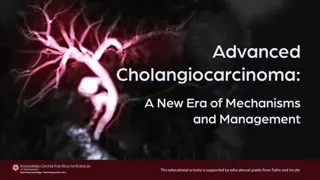
Cholangiocarcinoma Slow growing malignancy of biliary tract which tend to infiltrate locally and metastasize late 90% adenocarcinoma 60-70% at the bifurcation ofhepatic ducts 20-30% - in the distal CBD 5-10% - arise within the liver (peripheral)
Biliary Tree Neoplasms Clinical symptoms: Weight loss (77%) Nausea (60%) Anorexia (56%) Abdominal pain (56%) Fatigue (63%) Pruritus (51%) Fever (21%) Malaise (19%) Diarrheoa (19%) Constipation (16%) Abdominal fullness (16%). Symptomatic patients usually have advanced disease, with spread to hilar lymph nodes before obstructive jaundice occurs Associated with a poor prognosis
MRCP: Cholangiocarcinoma at the Bifurcation Klatskin tumour - Cholangiocarcinoma of junction of right & left hepatic ducts
Gallbladder Cancer 6th decade 1:3, Male:Female Highest prevalence in, Mexico, Chile, Japan, and Native American women. Risk Factors: 90% have gallstones, porcelain gallbladder polyps particulary larger than 10 mm, Choledochal cysts Uncommonly diagnosed preoperatively >80% with gallstones Clinical manifestation from abdominal pain to unexplained weight loss and jaundice Palpable RUQ mass Jaundice suggests local extention with ductal obstruction DS: US, EUS, CT Surgery is the only curative option : Treatment Tumors limited to the muscular layer (T1) need simple cholecystectomy
Adenocarcinomas is 80-90% Cancer spreads through the lymphatics, venous drainage, and with direct invasion Clinical Manifestations : vomiting. abdominal discomfort, right upper quadrant pain, nausea, and less common: Jaundice, weight loss, anorexia, ascites, and abdominal mass
Stent Placement -Endoscopic Approach A catheter is inserted through the endoscope into the ostium of the common bile duct. While maintaining the endoscope position in the duodenum, a wire is inserted through the catheter into the bile duct. The stent delivery system is then inserted over the wire to the site of obstruction, where the stent is deployed.
Bile Duct Carcinoma Etiology : Primary sclerosing cholangitis choledochal cysts ulcerative colitis hepatolithiasis anastomosis infection with Clonorchis or biliary-enteric in chronic typhoid carriers Adenocarcinomas is Over 95% Anatomically divided into distal, proximal, or perihilar tumors
Clinical Manifestations : Painless jaundice Pruritus, mild right upper quadrant pain, anorexia, fatigue, weight loss Diagnosis ultrasound CT scan : Cholangiography( PTC, ERC ) celiac angiography MRI Treatment : Surgical excision is the only potentially curative treatment unresectable perihilar cholangiocarcinoma need Roux-en- Y cholangiojejunostomy to segment II or III bile ducts or to the right hepatic duct
Complication History Examination Blood tests Biliary Colic - Intermittent RUQ/epigastric pain (minutes/hours) into back or right -Tender RUQ -No peritonism -Murphy s -WCC (N) CRP (N) - LFT (N) shoulder - N&V -Apyrexial, HR and BP (N) Acute Cholecystitis -Constant RUQ pain into back or right Tender RUQ - - - - WCC and CRP ( ) LFT (N or mildly ( ) - - shoulder -N&V -Feverish Periotnism RUQ (guarding/rebound) Murphy s + Pyrexia, HR ( ) Empyema -Constant RUQ pain into back or right Tender RUQ Peritonism RUQ Murphy s + - - - - - WCC and CRP ( ) LFT (N or mildly ( ) - - shoulder -N&V -Feverish Pyrexia, HR ( ), BP ( More septic than acute cholecystitis or ) Obstructive Jaundice Yellow discolouration Pale stool, dark urine - - - Jaundiced - - - - - WCC and CRP (N) - - ) - Non-tender or minimally tender RUQ LFT: obstructive pattern bili ( ), ALP ( ), GGT ( ), ALT/AST ( painless or assocaited with mild RUQ No peritonism Murphy s pain INR ( or ) Apyrexial, HR and BP (N) Ascending Cholangitis Becks triad -Jaundiced -Tender RUQ -Peritonism RUQ WCC and CRP ( ) - - ) - -RUQ pain (constant) LFT : obstructive pattern bili ( ), ALP ( ), GGT ( ), ALT/AST ( -Jaundice -Rigors Spiking high pyrexia (38-39) HR ( ), BP ( Can develop septic shock - - - INR ( or ) or ) Acute Pancreatitis Severe upper abdominal pain (constant) into back Profuse vomiting - -Tender upper abdomen -Upper abdominal or generalised -WCC and CRP ( ) -LFT: (N) if passed stone or obstructive pattern ifstone still in CBD - peritonism or ) -Usually apyrexial, HR ( ), BP ( Amylase ( ) - - INR/APTT (N) or ( ) if DIC Gallstone Ileus - 4 cardinal features of SBO -distended tympanic abdomen -hyperactive/tinkling bowel sounds
Acute Cholangitis Pus under pressure May be difficult to distinguish from acute cholecystitis Managed medically with support, antibiotics Drainage is key Etiology Stone disease Anomalous PBJ Malignant biliary obstruction Primary sclerosing cholangitis Post instrumentation Cholangiography Surgery Sphincterotomy Stents Charcot s triad Pyrexia, Pain, Jaundice Elevated liver enzymes Leukocystosis
