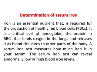
Triple Sugar Iron Test in Biochemical Analysis
The Triple Sugar Iron Test (TSI) is a crucial biochemical test used to differentiate gram-negative enteric bacilli based on their carbohydrate fermentation and hydrogen sulfide production. By examining color changes, gas production, and precipitate formation, microbiologists can interpret TSI results to identify specific bacterial characteristics. This test involves inoculating a TSI agar tube and incubating it to observe color changes and other reactions, with the results providing valuable insights into the metabolic capabilities of the tested bacteria.

Download Presentation

Please find below an Image/Link to download the presentation.
The content on the website is provided AS IS for your information and personal use only. It may not be sold, licensed, or shared on other websites without obtaining consent from the author.If you encounter any issues during the download, it is possible that the publisher has removed the file from their server.
You are allowed to download the files provided on this website for personal or commercial use, subject to the condition that they are used lawfully. All files are the property of their respective owners.
The content on the website is provided AS IS for your information and personal use only. It may not be sold, licensed, or shared on other websites without obtaining consent from the author.
E N D
Presentation Transcript
Biochemical Tests -2- 2022-2023
Head Lines in This Lecture Triple Sugar Iron Test Indole Test Urease Test Simmons Citrate Test
Triple Sugar Iron Test (TSI) Triple Sugar Iron Agar (TSI Agar) is used for the differentiation of gram-negative enteric bacilli based on carbohydrate fermentation and the production of hydrogen sulfide.
Triple Sugar Iron Test (TSI) Carbohydrate fermentation is detected by the presence of gas and a visible color change (from red to yellow) of the pH indicator, phenol red. The production of hydrogen sulfide is indicated by the presence of a precipitate that blackens the medium in the buttom of the tube.
Composition of Triple Sugar Iron Agar (TSI) 0.1% Glucose: If only glucose is fermented, only enough acid is produced to turn the buttom yellow. The slant will remain red. 1.0 % lactose/1.0% sucrose: a large amount of acid turns both buttom and slant yellow, thus indicating the ability of the culture to ferment either lactose or sucrose. Iron: Ferrous sulfate: Indicator of H2S formation Phenol red: Indicator of acidification (It is yellow in acidic condition and red under alkaline conditions). It also contains Peptone which acts as source of nitrogen. (when peptone is utilized under aerobic condition ammonia is produced)
Procedure for Triple Sugar Iron Agar (TSI) Test With a sterilized straight inoculation needle touch the top of a well-isolated colony Inoculate TSI Agar by first stabbing through the center of the medium to the bottom of the tube and then streaking on the surface of the agar slant. Incubate the tube at 37 C for 18 to 24 hours.
Interpretation of Triple Sugar Iron Agar Test If lactose (or sucrose) is fermented, a large amount of acid is produced, which turns the phenol red indicator yellow both in buttom and in the slant. Some organisms generate produces bubbles/cracks on the medium. gases, which
Interpretation of Triple Sugar Iron Agar Test If neither lactose/sucrose nor glucose is fermented, both the butt and the slant will be red. The slant can become a deeper red-purple (more alkaline) as a result of production of ammonia from the oxidative deamination of amino acids (peoptone is a major constituents of TSIAgar) . if H2S is produced, the black color of ferrous sulfide is seen.
Example of Triple Sugar Iron (TSI) Agar Reactions Name of the organisms Slant Butt Gas H2S Escherichia, Klebsiella, Enterobacter Acid (A) Acid (A) Pos (+) Neg (-) Shigella, Serratia Alkaline (K) Acid (A) Neg (-) Neg (- ) Salmonella, Proteus Alkaline (K) Acid (A) Pos (+) Pos (+) Pseudomonas Alkaline (K) Alkaline (K) Neg (-) Neg (-)
Indole Test This test demonstrate the ability of certain bacteria to decompose tryptophan to indole, which accumulates in the medium. Indole production test is important in the identification of Enterobacteria. Most strains of E. coli, P. vulgaris, and Providencia species break down the amino acid tryptophan with the release of indole. the amino acid
Indole Test This is performed by a chain of a number of different intracellular enzymes, a system generally referred to as tryptophanase. Tryptophan is an amino acid that can undergo deamination and hydrolysis by bacteria that express enzyme. tryptophanase
Indole Test When indole is combined with Kovac s Reagent (which contains hydrochloric acid and p-dimethylaminobenzaldehyde in amyl alcohol) the solution turns from yellow to cherry red. Because amyl alcohol is not water soluble, the red coloration will form in an oily layer at the top of the broth.
Procedure of Indole Test Take a sterilized test tubes containing 4 ml of tryptophan broth. Inoculate the tube aseptically by taking the growth from 18 to 24 hrs culture. Incubate the tube at 37 C for 24-28 hours. Add 0.5 ml of Kovac s reagent to the broth culture. Observe for the presence or absence of ring.
Result of Indole Test Positive: Formation of a pink to red color ( cherry-red ring ) in the reagent layer on top of the medium within seconds of adding the reagent. Aeromonas hydrophila, Aeromonas Bacillus alvei, Examples: punctata, Haemophilus influenzae, Proteus species. (not P. mirabilis and P. penneri), shigelloides, Pasteurella multocida, Pasteurella Enterococcus faecalis, and Vibrio species. Escherichia coli, pneumotropica,
Result of Indole Test Negative: No color change even after the addition of appropriate reagent. Actinobacillus Aeromonas Examples: salmonicida, most Bacillus spp., Bordetella spp., Enterobacter spp., Lactobacillus Haemophilus spp., Neisseria spp., Pasteurella Pasteurella ureae, Pseudomonas spp., Salmonella spp., Serratia spp., & Yersinia spp. spp., spp., most spp., Klebsiella most haemolytica, mirabilis, Proteus
Urease Test The determine organism to split urea, through the production of the enzyme urease and for the differentiation of enteric bacilli. urease test is used of to an the ability
Principle of Urease Test Urea is the product of decarboxylation of amino acids. Hydrolysis of urea produces ammonia and CO2. The formation of ammonia alkalinizes the medium, and the pH shift is detected by the color change of phenol red from light orange at pH 6.8 to pink at pH 8.1. Rapid medium pink within 24 hours. Weakly positive organisms may take several days, and negative organisms produce no color change or yellow as a result of acid production. urease-positive organisms turn the entire
Uses of Urease Test This identification of Proteus, Citrobacter Corynebacterium species. It is also useful to identify Cryptococcus spp., Brucella, Helicobacter other bacteria that produce the urease enzyme. Directly, this test is performed on gastric biopsy samples to detect the presence of H. pylori. test can of be several used as part and of species including the genera Enterobacteriaceae, Klebsiella, species, Yersinia as and some and some as well pylori, and many
Rapid Urease Test (RUT) The rapid urease test (RUT) is a popular diagnostic test for diagnosis of Helicobacter pylori. It is a rapid, cheap and simple test that detects the presence of urease in or on the gastric mucosa. It is also known as the CLO test (Campylobacter-like organism test). This test uses a gastric endoscopy and biopsy to collect stomach lining cells.
Simmons Citrate Test Simmons' citrate test is used for differentiating gram-negative bacteria on the basis of citrate utilization. Simmons' citrate agar is a defined, selective and differential medium that tests for an organism's ability to use citrate as a sole carbon source and ammonium ions as the sole nitrogen source.
Simmons Citrate Agar The medium contains citrate, ammonium ions, and other inorganic ions needed for growth. It also contains bromothymol blue, a pH indicator. Bromothymol blue is green at pH below 6.9, and then turns blue at a pH of 7.6 or greater.
Procedure of citrate utilization test Inoculate simmons citrate agar lightly on the slant by touching the tip of a needle to a colony that is 18 to 24 hours old. Incubate at 37oC for 18 to 24 hours. Some organisms may require up to 7 days of incubation due to their limited rate of growth on citrate medium. Observe the development of blue color; denoting alkalinization.
Results of citrate utilization test Citrate positive: growth will be visible on the slant surface and the medium color will change to blue. The alkaline bicarbonates produced as by-products of citrate catabolism raise the pH of the medium to above 7.6, causing the bromothymol blue to change from the original green color to blue . Klebsiella, Enterobacter Providencia, Proteus, Serratia, vibrio cholerae, Pseudomonas, Salmonella members of the subgenera Salmonella II, III and IV. carbonates and , Citrobacter, enteritidis and
Results of citrate utilization test Citrate negative: Trace or no growth will be visible. No color change will occur; the medium will remain the deep forest green color of the un inoculated agar. Yersinia enterocolitica. Salmonella Typhi. Escherichia coli. Salmonella Paratyphi A. Yersinia enterocolitica.
Reference Jawetz, Melnick, & Adelberg's Medical Microbiology, 28th Stefan Riedel, Jeffery A. Hobden, Steve Miller, Stephen A. Morse, Timothy A. Mietzner, Barbara Detrick, Thomas G. Mitchell, Judy A. Sakanari, Peter Hotez, Rojelio Mejia Originally published: August 25, 2019


")
")


 Agar")


")
")