Special Senses: Ears, Eyes, and Sound Waves
Explore the fascinating world of special senses including equilibrium, hearing, vision, and the science behind sound waves. Delve into the anatomy of the ears, inner cochlea, and trochlea, as well as what gives eyes their color. Discover how sound frequency and amplitude affect pitch and volume, and learn about the conjunctiva in the eyes.

Download Presentation

Please find below an Image/Link to download the presentation.
The content on the website is provided AS IS for your information and personal use only. It may not be sold, licensed, or shared on other websites without obtaining consent from the author.If you encounter any issues during the download, it is possible that the publisher has removed the file from their server.
You are allowed to download the files provided on this website for personal or commercial use, subject to the condition that they are used lawfully. All files are the property of their respective owners.
The content on the website is provided AS IS for your information and personal use only. It may not be sold, licensed, or shared on other websites without obtaining consent from the author.
E N D
Presentation Transcript
Special Senses The Ears (Equilibrium and Hearing) The Eyes (Vision)
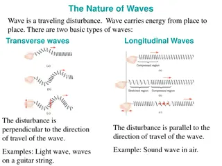
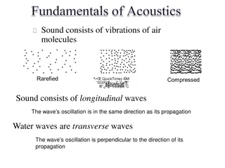
Sound Waves 1. Frequency = wave length -> pitch of sound 2. Amplitude = height of wave ->volume of sound
Conjunctiva - lines inside of eyelids and covers the sclera of the (the whites of the eye). Made of non-keratinized, stratified squamous epithelium with goblet cells, and stratified columnar epithelium. Accessory lacrimal glands in the conjunctiva produce the aqueous portion of tears. Additional cells present in the conjunctival epithelium include melanocytes, T and B cell lymphocytes. The conjunctiva can become inflamed due to an infection or an autoimmune response. This is known as conjunctivitis or referred to as pinkeye .
High Frequency Sound Low Frequency Sound
Vision Visual Conditions Myopia is called near (short) sightedness. Results in the light entering the eye not being directly focus on the fovea centralis of the retina, but rather in front of it. Causes distant image to be out of focus. Near focus not affected. Commonly from the eyeball being too long, alters distance of the fovea onto which the light is being refracted ( bent or focused). Myopia often treated by concave corrective lenses (have a negative optical power). Hyperopia is called far (long) sightedness causes the eyes to not have enough power to see close or nearby objects. Results in the light entering the eye not being directly focus on the fovea centralis of the retina, but rather in behind it. Hyperopia treated with convex corrective lenses (have a positive optical power).
Presbyopia = associated with aging in which the eye exhibits a progressively diminished ability to focus on near objects. Likely due to a decrease in elasticity of the lens and perhaps changes in the lens s curvature. Astigmatism - is caused by the shape of the cornea which prevents part of it from focusing light onto the retina. This results in a blurred area of the visual filed within an otherwise clear image. These conditions are termed "refractive errors as they involve changes in the way light is bent or focused as it enters the eye.
Normal Vision Light (image) focused on fovea of retina lens shape differs eye shape same Light focused behind fovea of the retina Lens is less flexible Light focused behind fovea of the retina Eye ball too short Light focused in front of fovea of the retina Eye ball too long