Skin Layers and Functions
Skin is composed of three main layers: the epidermis, dermis, and hypodermis. The epidermis serves as the protective barrier, while the dermis contains important structures like blood vessels and sweat glands. The hypodermis consists mainly of adipose tissue. Each layer plays a crucial role in functions such as protection, sensation, and thermoregulation. Different types of cells and layers within the skin contribute to its overall structure and function.

Download Presentation

Please find below an Image/Link to download the presentation.
The content on the website is provided AS IS for your information and personal use only. It may not be sold, licensed, or shared on other websites without obtaining consent from the author.If you encounter any issues during the download, it is possible that the publisher has removed the file from their server.
You are allowed to download the files provided on this website for personal or commercial use, subject to the condition that they are used lawfully. All files are the property of their respective owners.
The content on the website is provided AS IS for your information and personal use only. It may not be sold, licensed, or shared on other websites without obtaining consent from the author.
E N D
Presentation Transcript
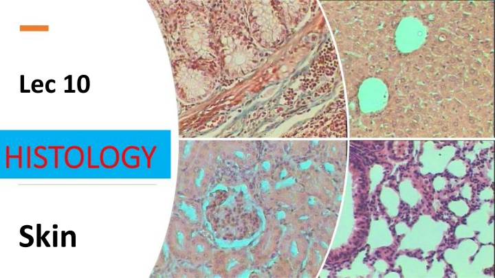
Lec 10 Skin
Three layers of skin The epidermis: a thin outer portion, that is the keratinised stratified squamous epithelium of skin. The important for the protective function of skin. The basal layers of this epithelium are folded to form dermal papillae. Thin skin contains four types of cellular layers, and thick skin contains five epidermis is
The dermis: a thicker inner portion. This is the connective tissue layer of skin. It is important for thermoregulation. It contains nerves, the blood supply, fibroblasts, etc, as well as sweat glands, which open out onto the surface of the skin, and in some regions, hair. The apical layers of the dermis are folded, to form dermal papillae, which are particularly prominent in thick skin. sensation, protection and The hypodermis. This layer is underneath the dermis, and merges with it. It mainly contains adipose tissue and sweat glands. The adipose tissue has metabolic functions: it is resonsible for production of vitamin D, and triglycerides.
The most superficial layer of theskin. The first barrier of protection from theinvasion of foreignsubstances. The epidermis is subdivided into five layers or strata: 1) Basal layer Stratum germinativeum. 2) Stratum spinosum (Malpighian layer). 3) Stratum Granulosum. 4) Stratum Lucidum. 5) Stratum Corneum.
(3) (3)- -Langerhans Langerhans cells: cells: They are (antigen-presenting cells) of theskin. Found in upper layers of st.spinosum. Have branched shape ¢ral nuclei. dendritic cells They keratin formation. Formed of many layers that continuously regenerate every 2-4 weeks. are responsible for immune shed and (2)-Melanocytes: (4)-Merkel cells Found in basal cell layer. They are sensory nervefibers. Function as touch receptors. Found inbetween cells of the basal layer. Branched centeral nuclei, organelles synthesizes (rER, Golgi, mitochondria). They form melanin. cells with contains protein for
Dermis: Thick skin has a thinner dermis than thin skin, and does not contain hairs, sebaceous glands, or apocrine sweat glands. There are only four layers in the epidermis of thin skin. The stratum lucidum layer is absent Dermis: Thin skin actually has a thicker dermis than thick skin, which makes thin skin easier to suture, if it gets damaged. Thin skin also has fewer eccrine/merocrine sweat glands. Thick skin is only found in areas where there is a lot of abrasion - fingertips, palms and the soles of your feet.
apoptosis Karyolysis is the complete dissolution of the chromatin of a dying cell due to the enzymatic degradation by endonucleases. The whole cell will eventually stain uniformly with eosin after karyolysis. It is usually associated with karyorrhexis and occurs mainly as a result of necrosis, while in apoptosis after karyorrhexis the nucleus usually dissolves into apoptotic bodies.


")