Role of Lipids in Plant-Pathogen Interaction
Lipids play a central role in signal molecule production for actively modifying the invasive capabilities of pathogens and the defensive mechanisms of plants during inter and intra-plant-pathogen interactions. They are essential constituents of membranes, providing structural support and serving diverse functions in energy storage, signal transduction, and stress responses. Lipid-modifying enzymes regulate the production of lipid-derived signaling molecules, influencing plant immunity and responses to microbial attacks. Key lipid classes involved include fatty acids, phospholipids, glycolipids, sterol lipids, sphingolipids, and waxes.
Download Presentation

Please find below an Image/Link to download the presentation.
The content on the website is provided AS IS for your information and personal use only. It may not be sold, licensed, or shared on other websites without obtaining consent from the author.If you encounter any issues during the download, it is possible that the publisher has removed the file from their server.
You are allowed to download the files provided on this website for personal or commercial use, subject to the condition that they are used lawfully. All files are the property of their respective owners.
The content on the website is provided AS IS for your information and personal use only. It may not be sold, licensed, or shared on other websites without obtaining consent from the author.
E N D
Presentation Transcript
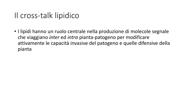
Il cross-talk lipidico I lipidi hanno un ruolo centrale nella produzione di molecole segnale che viaggiano inter ed intra pianta-patogeno per modificare attivamente le capacit invasive del patogeno e quelle difensive della pianta
Lipids Lipids are major constituents of prokaryotic and eukaryotic membranes. Besides serving as structural components of the plasma membrane and intracellular membranes, they provide diverse biological functions in energy and carbon storage, signal transduction, and stress responses Plants, fungi and bacteria contain a diverse set of lipids, including fatty acids, phospholipids, glycolipids, sterol lipids, sphingolipids, and waxes.
Lipids in host-pathogen interaction Membrane lipids are key players in plant cells during the response to microbial attack and during interactions with beneficial microbes. The expression of several genes encoding enzymes of lipid metabolism is upregulated after infection of plant cells, resulting in the synthesis, modification, or re-allocation of lipid-derived molecules. Lipid-modifying enzymes are essential regulators of the spatial and temporal production of lipid metabolites involved in signaling and membrane proliferation for the establishment of intracellular compartments or compositional changes of lipid bilayers Phospholipids and phospholipases Glycolipids Free fatty acids Glycerolipid Oxylipins Sterol lipids Carotenoids and apocarotenoids Sphingolipids
Phospholipids and phospolipases Phospholipids contain two fatty acids esterified to the sn-1 and sn-2 positions of a glycerol backbone, and a polar headgroup attached to the sn-3 position. Phospholipids of plants mainly comprise phosphatidic acid (PA), phosphatidylserine (PS), phosphatidylcholine (PC), phosphatidylethanolamine (PE), phosphatidylglycerol (PG), and phosphatidylinositol (PI). Each phospholipid class includes many molecular species due to a large number of fatty acids varying in chain length and degree of desaturation. Phospholipids are predominantly synthesized at the endoplasmic reticulum (ER). Phospholipases and phospholipid-derived molecules are involved in signaling and plant immunity during plant pathogen interactions. Phospholipases catalyze the conversion of phospholipids into fatty acids and lysophospholipids, diacylglycerol (DAG), or PA, depending on their positional specificity Upon microbe infestation, phospholipid-hydrolyzing enzymes are activated, contributing to the establishment of an appropriate defense response by inducing the production of defense-signaling molecules such as oxylipins, including jasmonic acid (JA), and the potent second messenger PA PLD PA PLA FA and LPE/C/G PLC DAG and IP3
Phospholipids and phospolipases Phospholipase D (PLD) cleaves the terminal phosphodiester bond of phospholipids, resulting in the formation of PA. PLDs have diverse functions in lipid metabolism and hormone signaling (abscisic acid, ABA; JA) and during responses to biotic and abiotic stress. PLDs and PLD-derived PA play important roles in the plant defense response PLD-derived PA often directly binds to proteins, leading to alterations in protein localization or enzyme activity. PLD-derived PA targets more than 30 proteins involved in diverse physiological pathways PA regulates a range of physiological processes such as the activity of kinases, phosphatases, phospholipases, and proteins involved in membrane trafficking, Ca2 +signaling, or the oxidative burst. PA serves as the precursor for the lipid intermediates LPA, DAG, and free fatty acids, all of which can be involved in plant defense signaling
Phospholipids and phospolipases The group of phospholipases C (PLC) in plants can be divided into three families according to substrate specificity and cellular function: (i) PC-PLCs or non-specific PLCs that hydrolyze PC and other phospholipids, (ii) phosphatidylinositol-4,5-bisphosphate (PIP2)-PLCs (PI-PLC) that act on phosphoinositides, and (iii) glycosylphosphatidylinositol (GPI-PLCs) that hydrolyze GPI anchors on proteins. PI-PLCs cleave PIP2, producing DAG and IP3(1,4,5-inositol trisphosphate) both acting as second messengers. PI-PLC activity is stimulated in plants in response to pathogenic infection. PAMP recognition triggers the activation of the PLC/DAG kinase pathway, resulting in the accumulation of PA. Thus, PA in part originates from DAG produced by PLC because DAG can be further phosphorylated by DAG kinase (DGK) PLC does not only play a role in elicitor recognition processes, but also in downstream disease resistance signaling
Phospholipids and phospolipases The phospholipase A (PLA) superfamily is divided into PLA1and PLA2families. PLA enzymes catalyze the hydrolysis of the acyl ester bonds of phospholipids at their sn-1 and sn-2 positions, respectively, yielding free fatty acids and lysophospholipids. PLA2-derived LPC and LPE are involved in systemic responses after wounding. Lysophospholipids are further hydrolyzed by lyso-PLAs yielding glycerophosphodiesters. PLAs are believed to be involved in the regulation of plant growth, root and pollen development, stress responses, and defense signaling. They have been mainly linked to plant immunity through their role in oxylipin and JA biosynthesis and the stimulation of downstream defense products The enzymatic products of PLA and lyso-PLA activities are glycerophosphodiesters, which are further hydrolyzed by glycerophosphodiester phosphodiesterases yielding Gro3P (Glycerol-3-phosphate). Gro3P accumulation is a highly conserved process in different organisms
Glycolipids & SAR Glycolipids are abundant membrane components in chloroplasts of plants and algae and in cyanobacteria, and some bacterial phyla. Galactolipids make up the major glycolipid fraction in plants because monogalactosyldiacylglycerol (MGDG) and digalactosyldiacylglycerol (DGDG) Galactolipids play important roles in signal transduction, cell communication and pathogen responses. The two galactolipids apparently have different functions in SAR because MGDG regulates the biosynthesis of AzA and Gro3P, while DGDG affects the biosynthesis of NO and SA and is also required for AzA - induced SAR in Arabidopsis
Ossilipine come segnali di sviluppo e comunicazione ospite-patogeno I patogeni e i loro ospiti hanno acquisito dei complessi meccanismi di signallingper interagire con l ambiente circostante Le ossilipine rappresentano una nuova classe di molecole implicate nel signalling ospite-patogeno Le ossilipine rappresentano una famiglia molto diversificata di metaboliti secondari che si originano dall ossidazione o da un ulteriore bio-conversione degli acidi grassi polinsaturi (PUFA) e monoinsaturi (UFA)
Le ossilipine Le ossilipine includono gli idroperossi-, idrossi-, oxo- ed epossi-acidi grassi, divinil-eteri, aldeidi volatili e jasmonati Le ossilipine possono formare coniugati esterificati come i glicerolipidi (glicolipidi, fosfolipidi, e lipidi neutrali) Le ossilipine possono avere diversi ruoli biologici: Secondi messaggeri Antimicrobici Insetticidi Antifungini
Le pathways di biosintesi delle ossilipine Le ossilipine sono sintetizzate o per ossidazione chimica (radicalica) oppure enzimaticamente dai PUFA da tre pathways principali La lipossigenasi (LOX) Le alfa-diosigenasi ( -DOX) (piante) e linoleato diolo sintasi (LDS) (funghi) che mostrano similarit con le COX animali e catalizzano la -ossidazione degli FA Attraverso gli enzimi del citocromo P450 (CYP74) localizzati nell ER che catalizzano la o in chain-hydroxylation degli acidi grassi
Non-enzymatic lipid peroxidation La perossidazione non enzimatica dei lipidi di membrana catalizzata durante lo stress ossidativo e la formazione di specie reattive dell ossigeno (ROS) come il perossido d idrogeno e l ossigeno singoletto L effetto tossico dei ROS pu essere causato principalmente dalla loro conversione nell idrossile radicale (HO ) specie radicalica altamente reattiva. Questo radicale attacca i lipidi di membrana e porta all ossidazione dei PUFA pi abbondanti presenti nelle membrane delle cellule vegetali, cio l acido linoleico e linolenico La miscela racemica risultante dei radicali perossidici dei PUFA pu , a sua volta, iniziare una reazione a catena che porta alla formazione e all accumulo di racemi di idroperossidi di acidi grassi che sono prevalentemente esterificati in complessi lipidici nelle membrane oppure permangono liberi nel citoplasma
LOX-oxylipin biosynthesis Le LOX sono classificate in base alla loro regio-specificit cio in base alla posizione in cui introducono l ossigeno molecolare nel PUFA. L ossidazione del C18:2 ad esempio pu avvenire sia al carbonio 9 (9-LOX) che al C13 della catena alifatica Le LOX possono utilizzare come substrato sia degli acidi grassi liberi che complessati in fosfolipidi o trigliceridi I geni che codificano per LOX sono presenti come famiglie mutligeniche in tutte le piante analizzate, ad es. sono 6 in Arabidopsis thaliana e 14 in patata; 2 in Aspergillus flavus; 4 in A. ochraceus
Enzymatic oxylipin biosynthesis Il metabolismo dei LOX-derived hydroperoxy fatty acids avviene attraverso 6 vie alternative Quattro di queste sono catalizzate da un famiglia atipica di citocromo P450 monossigenasi, gli enzimi CYP74: Allene ossido sintasi Idroperossido liasi Divinil etere sintasi Epossi alcool sintasi
9 e 13 ossilipine I principali prodotti perossidati derivanti dai 13- idroperossidi (13-HPOD/TE) della AOS sono un gruppo di composti ciclopentanonici (se derivano dal C18:3) chiamati jasmonati e che includono l acido jasmonico (JA), il metil jasmonato (MeJA) e l acido 12-oxo-fitodienoico (12-OPDA) Questi composti sono implicati nell hormone signalling scatenato dalla risposta a stress biotici e abiotici I prodotti del ramo HPL sono i cosiddetti green leaf volatiles (GLVs). Ad es. lo Z-3-esenale, dovrebbe avere una funzione come regolatore della difesa od avere un attivit antifungina
9 e 13 ossilipine La sintesi dei divinil eteri (DE) ristretta ad alcune specie vegetali e le diverse isoforme di DES catalizzano la formazione di una moltitudine di prodotti I DE potrebbero avere un ruolo nelle reazioni di difesa in tubero di patata contro patogeni come la Phytophthora infestans Meno caratterizzati sono i prodotti della EAS. Tra questi il pi conosciuto anche per applicazioni biotecnologiche l acido vernolico (acido 12,13-epoxy-octadec-cis-9-enoic) che presente nei semi di Crepispalaestina Questo epossido comunque coinvolto con la sintesi di suberina e cutina anche durante le reazioni di stress
-DOX/LDS oxylipin biosynthesis L -ossidazione catalizzata dalle -DOX porta alla formazione di un derivato instabile, (2R-idroperossido), di un acido grasso (UFA o PUFA). Questo rapidamente convertito in un corrispondente -idrossi acido grasso, in aldeidi a corta catena cos come in acidi grassi a corta catena LDS1 (linoleato diolo sintasi)trasforma il 18:2 (acido linoleico) in ossilipine come 8R-HPODE e di-HODE
La percezione delle ossilipine Le ossilipine fungine non vengono stoccate in compartimenti ma sintetizzate de novo dai C18 insaturi quando le cellule sono attivate da stimoli interni o esterni. Le ossilipine escono dalle cellule attraverso degli specifici ABC transporter e funzionano localmente a bassi livelli (concentrazioni nM) attraverso processi autocrini o paracrini (cio segnalano nel sito di sintesi o nelle immediate vicinanze) sui recettori superficiali legati a G protein Un GPCR di mammifero (G2A) funziona come recettore per il 9-HODE L attivazione dei GPCR porta a cambiamenti nei componenti prossimali delle receptor-mediated pathways (cio G protein, Ras) che attivano dei componenti intermedi di signalling (es. fosfolipasi C, PkaA e PKCs) che alla fine alterano i livelli di cAMP o di ione calcio che servono come secondi messaggeri che stimolano i meccanismi di signaling alla base di molti processi cellulari
Sterol lipids Sterol lipids are synthesized via the isoprenoid pathway in the cytosol of plant cells. Sterols can occur in their free form (free sterols) or are derivatized at the C3 hydroxy group (conjugated sterols). The result of aggregation of sterol lipids in the plasma membrane, form microdomains (lipid rafts) enriched in sterol lipids and specific plasma membrane resident proteins.
Plant sterol lipids Free sterols (FS) and the conjugated forms of sterol glucosides (SG) and acylated sterol glucosides (ASG) are constituents of extraplastidial membranes, while sterol esters (SE) are deposited in oil bodies in the cytosol. The increase in resistance of the Arabidopsis CYP710A1 mutant is attenuated after exogenous stigmasterol application, while - sitosterol application had the opposite effect. These results show a correlation between an increased stigmasterol/ -sitosterol ratio and bacterial virulence, probably due to changed membrane integrity resulting from the shift in sterol lipids P. syringae induced accumulation of stigmasterol in membranes represents a defense response of plants to prevent unwanted nutrient efflux and thus bacterial proliferation in the apoplast
Fungal sterol lipids Ergosterol is the most abundant sterol in most fungi, and it is therefore employed as fungal lipid marker. It is one of the MAMPs that acts as elicitor of microbe-triggered immunity (MTI) upon contact with the plant. Lanosterol derived from oxido-squalene is an important intermediate in the synthesis of sterols in plants and fungi. However, the pathway of sterol synthesis from lanosterol differs between plants and fungi. The final step of ergosterol synthesis in fungi is the conversion of ergosta-5,7,22,24(28)-tetraenol to ergosterol catalyzed by the C-24 reductase ERG4. Fungal erg4 mutants of Fusarium graminearum are devoid of ergosterol. The erg4 mutants were capable of plant colonization but showed reduced virulence, which was attributed to reduced mycelia growth, increased sensitivity to ROS due to impaired membrane integrity and a decrease in deoxynivalenol content, a toxin essential for aggressiveness
Sterylglycosides SGs are sugar derivatives of a membrane-bound sterol. The sterol consists mainly of sitosterol, sigmasterol, and campesterol in plants, ergosterol in fungi, and cholesterol in animals. SGs are characterized by a planar sterol backbone made up of four condensed aliphatic rings and a hydrocarbon side chain at C17 with the sugar moiety attached to the 3 -hydroxy group at carbon 3 of the sterol. Studies on the role of SGs have shown that these glycolipids are important regulators of the host immune response to fungal infections Sterylglycosyltransferases and sterylglucosidases have been identified in fungi, yeast and plants. In fungi they act as virulence factors The Sgl1 enzyme is an important virulence factor that controls the intracellular fungal levels of SGs in order to prevent a host immune response against the pathogen
Sphingolipids Ceramide (Cer) is the fundamental unit of all complex sphingolipids. The Cer core consists of two structural moieties: the sphingoid long- chain base (LCB) and the fatty acid (FA) chain linked via an amide bond. The typical LCB has a chain length of 18 carbons, which may be hydroxylated at C4 (1), or have a double bond at the 4 or 8 carbon (2). The FA chain may be hydroxylated at the - position (3) , and/or have a double bond at 9- position (4).The FA chain length may vary from 14 to 36 [if >20, it is referred to as very long- chain FA, i.e., VLCFA] (5). The structurally diverse ceramides can be converted to more complex sphingolipids via substitution of the head group designated R at the 1-position of the LCB (6). Additional sugar residues may be further added to IPCs and GlcCERs, resulting in more complex sphingolipids. the four major classes of plant sphingolipids are free LCBs, ceramides, glycosylinositol phosphoceramides (GIPC) glucosylceramides (GlcCer)
Gli Sfingolipidi come e dove si producono Complex sphingolipids can be formed via two major pathways: the de novo biosynthesis pathway, starting with the condensation of a serine with an acyl-CoA; the salvage pathway, where ceramides and LCBs as catabolites of more complex sphingolipids re-enter the synthetic pathway
La sfingobiologia Starting at top left, serine and palmitoyl-CoA are condensed by serine palmitoyltransferase (SPT) to form 3-ketosphinganine that is reduced to sphinganine: N-acylated by ceramide synthases (CerS) with the shown fatty acyl-CoA preferences, or Phosphorylated by sphingosine kinase (SphK). The N-acylsphinganines (dihydroceramides, DHCer) are also oxidized to Cer by dihydroceramide desaturase (DES1 and DES2). DES2 is also capable of hydroxylating the 4-position to form 4-hydroxydihydroceramides, (t18:0) and incorporated into more complex sphingolipids as shown. For example, d18:1/C16:0 refers to a ceramide consisting of dihydrosphingosine with an 18 carbon chain plus 1 double bond (d18:1), and an amide-linked C16 FA chain with 0 double bond (C16:0).
Gli Sfingolipidi cosa fanno GlcCer and GIPC are abundant components of the plasma membrane, tonoplast, and ER membrane in plant cells and together with sterols, they are involved in membrane rafts formation Arabidopsis ceramide synthases (LOH 1-3) display substrate specificity toward specific LCBs and fatty acyl-CoAs LOH2 is specific for the synthesis of ceramides containing a dihydroxy LCB and C16 fatty acid (e.g., d18:1-h16:0), whereas LOH1 and LOH3 synthesize ceramides with a trihydroxy LCB and VLCFAs (e.g., t18:1- h24:0). Apparently, the resulting ceramides have very distinct functions in cell metabolism.
Sphingolipids and plant immunity Since the identification of protein kinase C as the first target of sphingosine in mammalian cells, bioactive sphingolipids have been shown to target many proteins and regulate their cellular functions in yeast and animals. The mitogen-activated protein kinase 6 (MPK6) represents a potential direct protein target downstream of LCBs in the signal transduction leading to cell death (PCD). (i). MPK6 is a known component of the MAP kinase signaling module of PTI (ii). MPK6 also contributes to ETI and SA-dependent basal (iii). MPK6 is targeted and inhibited by HopA1, an effector protein of P. syringae and (iv). t18:0 is rapidly accumulated before the onset of HR and ETI Plants develop ETI in part through reinforcing PTI, and activation of MPK6 by bioactive free LCBs de novo synthesized in infected tissues during early signaling of ETI may act as a bridge connecting the ETI and PTI pathways. MPK6 is also activated during immune response induced by pathogen-specific sphingolipids which presumably function as PAMPs
Lipid rafts Membrane fractions characterized by a particular lipid composition leading to a lo or highly-organized phase operationally defined in terms of insolubility in non- ionic aqueous detergents The components of biological membranes can be arranged in a nonhomogeneous lateral distribution, leading to the creation of ordered structures that differ in lipid and/or protein composition from the surrounding membrane These structures are believed to arise from preferred interactions between saturated lipids, glycosphingolipids, sphingomyelin, and sterols that give rise to a sterol-dependent liquid ordered phase (Lo) which can coexist with a liquid disordered (Ld) phase under physiological conditions They are mobile, dynamic entities that move laterally along the plane of the plasma membrane and traffic continuously between the plasma membrane and internal compartments These membrane regions play crucial roles in signal transduction, phagocytosis and secretion, as well as pathogen adhesion/interaction.
Lipid rafts basic constituents In mixtures comprised of bilayer forming lipids such as dipalmitoyl-PC and cholesterol (or ergosterol in yeast), a third physical phase, the liquid-ordered (lo) phase can be observed. In the lo phase, the acyl chains of lipids are extended and ordered, as in the gel phase, but have higher lateral mobility in the bilayer. Sterols can stabilize the lo phase by filling in the hydrophobic gaps between the phospholipid or glycolipid acyl chains SL are ceramide-based amphipathic lipids that can create a complex network of hydrogen bonds because of the presence in the ceramide moiety of amide nitrogen, carbonyl oxygen, and a hydroxyl group positioned near the water/lipid interface of the bilayer the geometrical properties resulting from the bulky hydrophilic head group of GSLs strongly favor phase separation and spontaneous membrane curvature The association of sterols with sphingolipids promotes phase separation apparently because of favourable packing interactions between saturated lipids and sterol
Lipid-protein interaction in the rafts Protein and lipid-driven lateral organization have been regarded as somehow mutually exclusive, but it has become clear that they cooperate in the creation of structural and functional heterogeneity in membranes. Some proteins that are associated with lipid rafts are surrounded by a shell of typical raft lipids (SLs, sterol). Such a shell may confer to a membrane protein a higher affinity for lipid rafts, resulting in its partitioning to a phase-separated membrane domain in cooperation with or even in the absence of a specific raft targeting motif. Sterol-binding domains and SL-binding domains (that bind to the polar head groups of SLs) have been identified and characterized in several proteins. The binding of lipids to receptors induces conformational changes that affect both ligand binding and signaling pathways downstream of receptor activation.
Which proteins in the rafts? Lipid-to-receptor binding also influences the lateral organization of membrane components in the domain of a membrane-organizing protein. Several classes of membrane-associated proteins display a strong association with lipid rafts. A common raft-targeting motif is the presence of a GPI anchor, which is sometimes modified by acylation of the inositol head group or by replacement of the glycerolipid residue with ceramide Certain raft-associated proteins appear to play important roles in organizing multiprotein complexes within lipid rafts These close-packed, highly saturated rafts have different properties compared with the surrounding, less ordered and highly unsaturated phospholipid bilayer. Due to these different properties certain transmembrane and membrane-associated proteins preferentially insert into these rafts. In mammalian cells post- translational modifications such as myristoylation and palmitoylation were shown to target proteins preferentially to membrane rafts in the cytoplasmic membrane leaflet, while the addition of a glycosylphosphatidylinositol (GPI) moiety anchors proteins pre- dominantly to membrane rafts at the outer leaflet
Knowledge from animals lipid rafts and signaling in T cells T lymphocytes membranes were characterised as large non-covalent complexes containing GPI-anchored proteins and Src family tyrosine kinases that can transduce activation signals to internal Src family kinases T cell antigen receptor (TCR) engagement triggers the assembly of a large macromolecular complex containing a variety of signalling molecules and adapters, actually rafts are the platforms for this signalling complex In resting T cells, rafts are highly enriched in the Src kinases Lck and Fyn and the linker for activation of T cells (LAT) transmembrane adapter. Extensive crosslinking of the TCR with antibodies promotes the rapid activation of Src kinases and subsequent accumulation in rafts of a series of newly tyrosine phosphorylated substrates Upon assembly of the signalling machinery, the cytoskeleton is reorganised, and the Ras/MAPK and PLCg1 cascades are activated within the rafts, which produce signals that stimulate T cell proliferation
Knowledge from animals lipid rafts and membrane traficking in T cells In MDCK epithelial cells, newly synthesised proteins are segregated after passage through the Golgi in different vesicular carriers destined for the apical (red) and basolateral (green) subdomains Partitioning of proteins into rafts appears to mediate the sorting of at least some apical membrane proteins Caveolae are raft containing invaginated structures exclusively located in the basolateral surface. MAL and caveolin-1 are machinery involved in raft-dependent apical transport (straight arrow in red) and caveolae formation (curved arrow in red), respectively Specifically, caveolin-1 is necessary for caveolae formation and organises lipid rafts to build the caveolar architecture. (D) MAL is necessary for apical transport and appears to organise lipid rafts for the formation of the transport vesicles.
Lipid rafts during immune responses The ability to dynamically reorganize the protein content of the plasma membrane is essential for the regulation of processes such as polarity of transport, development, and microbial infection. While the plant cell wall represents the first physical and mostly unspecific barrier for invading microbes, the plasma membrane is at the forefront of microbial recognition and initiation of defense responses.
Lipid rafts during plant immune responses In planta evidence for dynamic compartmentalization of membrane proteins was also reported upon host cell infection of Hordeum vulgare (barley) and A. thaliana by the powdery mildew fungus Blumeria graminis. Focal accumulations around fungal entry sites were observed for three otherwise evenly distributed fluorescently labeled proteins all involved in powdery mildew penetration resistance, namely MILDEW-RESISTANCE- LOCUS-O (MLO), the syntaxin HvREQUIRED-FOR-MLO-SPECIFIED- RESISTANCE- 2 (HvROR2) and its ortholog AtPENETRATION-1 (AtPEN1) A possible membrane raft localization of NtRBOHD is also supported by its identification in DIMs from N. tabacum after treatment with cryptogein, together with its negative regulator RAC/ROP GTPase NtRAC5
Lipid rafts as entry points for intracellular microbials The integral membrane proteins FLOTILLIN-1 and FLOTILLIN- 2 (synonymously called REGGIE-2 and REGGIE-1) are frequently used as markers for membrane rafts in the cytoplasmic leaflet of mammalian cells Members of this protein family were described to be involved in clathrin-independent endocytosis and are present on host-derived membranes surrounding intracellular microorganisms, indicating raft-mediated endocytosis as a possible entry point for microorganisms Furthermore, evidence that flotillins are interacting and/or co-localizing with many signaling components, such as receptors and mitogen- activated protein kinases (MAPK), suggests that these proteins may act as scaffolds for a number of signaling processes Membrane rafts serving as signaling hubs that can be exploited by (facultative) intracellular microbes to successfully establish themselves in their host.
Pathogens and rafts: interacting to survive An interesting manner that allows pathogens to evade the immune system is through membrane microdomains. As signalling for the innate and adaptative immune responses is initiated in rafts, some pathogens have evolved mechanisms to subvert this signalling by co- opting raft-associated pathways Several pathogens can directly interact with different target cells through membrane microdomains in different manners. It is known that the entry via lipid rafts can avoid lysosomal fusion and therefore allow pathogen survival. In addition, parasites might modulate signalling pathways, including lipid-raft-associated protein kinases [Srfk (Src family kinases)]. Especially viruses, which do not have their own protein synthesis machinery, target the ER after subverting the lysosomal pathway
Lipid signals in plant-pathogen interaction: a case of study Fusarium verticillioides Zea mays 9-oxylipins produced by maize caryopses in response to fungal attack, trigger the synthesis of FB1 FB1 synthesis increase is closely modulated at chromatin level since oxylipins trigger the hyperacetylation of H4 at the promoter site of fum1, a gene involved in FB1 synthesis In turn, FB1 produced hugely during the last stage of kernel development should hamper the activity of ceramide synthases into maize kernels
Interaction model Early stages of infection Late stages of infection F. verticillioides spend the early stages of infection as an endophytic, parasitic, symbiont in the aleurone layer of kernels. As kernel maturation occurs, it switches to necrotrophic behaviour FB1 could represent a signal for PCD since its accumulation block ceramide synthase and as reported elsewhere the augmentation of LCB d/t18:1-C16:0 may enhance the activity of MPK6 upstream to SA accumulation and providing a signal for PCD onset
