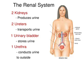
Renal Disorders and Function Tests
This resource delves into crucial functions of kidneys, renal glomerular function, renal tubular function, and the biochemistry of renal disorders. It explores topics such as excretory function, homeostasis, endocrine functions, and metabolic function related to renal health.

Download Presentation

Please find below an Image/Link to download the presentation.
The content on the website is provided AS IS for your information and personal use only. It may not be sold, licensed, or shared on other websites without obtaining consent from the author.If you encounter any issues during the download, it is possible that the publisher has removed the file from their server.
You are allowed to download the files provided on this website for personal or commercial use, subject to the condition that they are used lawfully. All files are the property of their respective owners.
The content on the website is provided AS IS for your information and personal use only. It may not be sold, licensed, or shared on other websites without obtaining consent from the author.
E N D
Presentation Transcript
Renal disorders and renal function test By Dr.Qutaiba Abdulkareem Qasim 2020
Topics to be discussed 1-Renal function : Renal glomerular function Renal tubular function Water reabsorption: urinary concentration and dilution 2-Biochemistry of renal disorders : Acute and chronic kidney injury Investigation of acute renal dysfunction Nephrotic syndrome Nephritic syndrome 3-Diagnosis of renal dysfunction
Crucial functions of kidneys 1-Excretory function : excretion of metabolic waste products , medications and toxic substances . 2-Homeostatic function : Control the body solute and water status , acid base balance. 3-Endocrine functions : -Production of 1,25-dihydroxyvitamin D, the active metabolite of vitamin D, which is produced following hepatic hydroxylation of 25-hydroxyvitamin by the renal enzyme 1, -hydroxylase, -Production of erythropoietin, which stimulates erythropoiesis. -Production of renin essential for blood pressure regulation 4-Metabolic function : gluconeogenic organ in prolonged fasting
RENAL GLOMERULAR FUNCTION About 200 L of plasma ultrafiltrate usually enter the tubular lumina daily, mainly by glomerular filtration . ( why this huge volume filterd although urine output only 0.8-2L /day?) Production of ultrafiltrate depends on the blood flow through normal glomeruli and on the difference between the hydrostatic pressure gradient and the plasma effective colloid osmotic (oncotic) pressure gradient across the membranes and tight junctions. The colloid osmotic effect is weak relative to the hydrostatic gradient but does facilitate some reabsorption of fluid from the proximal renal tubules. The filtrate contains diffusible constituents at almost the same concentrations as in plasma.
RENAL TUBULAR FUNCTION Reclamation of solute from the proximal Tubule : Almost all the potassium is actively reabsorbed from the proximal tubules, as is more than 70 per cent of the filtered sodium, free ionized calcium and magnesium. This reabsorption may be stimulated by parathyroid hormone (PTH) and inhibited by loop diuretics such as furosemide. Bicarbonate is almost completely recovered following exchange of sodium and hydrogen ions . Specific active transport mechanisms result in the almost complete reabsorption of glucose, urate and amino acids.
RENAL TUBULAR FUNCTION Phosphate reabsorption is incomplete; phosphate in tubular fluid is important for buffering hydrogen ions. Inhibition of phosphate reabsorption by PTH occurs in both the proximal and the distal convoluted tubules, and accounts for the hypophosphataemia of PTH excess. Almost all the filtered metabolic waste products, such as urea and creatinine, which cannot be reused by the body, remain in the luminal fluid. almost all the reusable nutrients and the bulk of electrolytes are reclaimed from the proximal tubules
WATER REABSORPTION: URINARY CONCENTRATION AND DILUTION Two main processes are involved in water reabsorption: Isosmotic reabsorption of water from the proximal tubules. The nephrons reabsorb 99 per cent of the filtered water, about 70 80 per cent (140 160 L/day) of which is returned to the body from the proximal tubules. Active solute reabsorption from the filtrate is accompanied by passive reabsorption of an osmotically equivalent amount of water. Therefore, fluid entering the lumina of the loops of Henle, although much reduced in volume, is still almost isosmotic. Dissociation of water reabsorption from that of solute in the loops of Henle, distal tubules and collecting ducts. Normally between 40 and 60 L of water enter the loops of Henle daily. This volume is reduced to about 2 L as varying amounts of water are reabsorbed, helping to correct for changes in extracellular osmolality. At extremes of water intake, urinary osmolality can vary from about 40 to 1400 mmol/kg. The proximal tubules cannot dissociate water and solute reabsorption, and the adjustment must occur between the end of the proximal tubule and the end of the collecting duct.
Mechanisms of water reabsorption in loops of Henli and collecting ducts Two mechanisms are involved: Countercurrent multiplication is an active process occurring in the loops of Henle, whereby a high osmolality is created in the renal medulla and urinary osmolality is reduced. This can occur in the absence of antidiuretic hormone (ADH), also called arginine vasopressin or vasopressin, and a dilute hypo-osmolal urine is produced. Countercurrent exchange is a passive process, occurring only in the presence of ADH. Water without solute is reabsorbed from the collecting ducts into the ascending vasa recta along the osmotic gradient created by countercurrent multiplication and by the high osmolality in the medulla, producing a concentrated urine.
Water restriction By increasing the plasma osmolality, water restriction increases ADH secretion and allows countercurrent exchange with enhanced water reabsorption. Reduced circulatory volume results in a sluggish blood flow in the vasa recta and increased urea reabsorption, allowing a build-up of the medullary hyperosmolality produced by multiplication. This potentiates water reabsorption in the presence of ADH. The reduced capillary hydrostatic pressure and increased colloid osmotic pressure, due to the haemoconcentration following water restriction , ensure that much of the reabsorbed water enters the vascular compartment.
Water load A high water intake dilutes the extracellular fluid, and the consequent fall in plasma osmolality reduces ADH secretion. The walls of the collecting ducts therefore remain impermeable to water and the countercurrent multiplication produces a dilute urine and a high osmolality within the medulla and medullary vessels. Blood from the latter flows into the general circulation, so helping to correct the fall in systemic osmolality. Increasing the circulating volume increases renal blood flow; the rapid flow in the vasa recta washes out medullary hyperosmolality, returning some of the solute, without extra water, to the circulation. Thus, not only is more water than usual lost in the urine, more solute is reclaimed .
Osmotic diuresis An excess of filtered solute in the proximal tubular lumina impairs the bulk water reabsorption from this site by its osmotic effect. The resulting impairment of build-up of medullary osmolality inhibits distal water reabsorption, under the influence of ADH from the collecting ducts, resulting in a diuresis . For example, glucose and urea are sometimes filtered at high enough concentration to exceed the proximal tubular reabsorptive capacity. They can then act as osmotic diuretics and cause water depletion. This is important, for example, in diabetes mellitus or in uraemia. The most effective osmotic diuretics are substances that cannot cross cell membranes to any significant degree; therefore, they must be infused, as they cannot be absorbed from the gut. One example is mannitol, a sugar alcohol, which is sometimes used therapeutically as a diuretic.
Homeostatic solute adjustment in the distal tubule and collecting duct Sodium reabsorption in exchange for hydrogen ions occurs throughout the nephrons. In the proximal tubules the main effect of this exchange is on reclamation of filtered bicarbonate. In the distal tubules and collecting ducts, the exchange process is usually associated with net generation of bicarbonate to replace that lost in extracellular buffering. Potassium and hydrogen ions compete for secretion in exchange for sodium ions. The possible mechanism stimulated by aldosterone . The most important stimulus to aldosterone secretion is mediated by the effect of renal blood flow on the release of renin from the juxtaglomerular apparatus; this method of reabsorption is part of the homeostatic mechanism controlling sodium and water balance.
BIOCHEMISTRY OF RENAL DISORDERS Pathophysiology : Different parts of the nephrons are in close anatomical association and are dependent on a common blood supply. Renal dysfunction of any kind affects all parts of the nephrons to some extent, although sometimes either glomerular or tubular dysfunction is predominant. The net effect of renal disease on plasma and urine depends on the proportion of glomeruli to tubules affected and on the number of nephrons involved. Uraemia is the term used to describe a raised plasma urea concentration and is almost always accompanied by an elevated creatinine concentration: in North America this is usually referred to as azotaemia (a raised nitrogen concentration).
Reduced glomerular filtration rate with normal tubular function Plasma High urea (uraemia) and creatinine concentrations.? Low bicarbonate concentration, with low pH (acidosis).? Hyperkalaemia.? Hyperuricaemia and hyperphosphataemia.? Urine Reduced volume (oliguria). Low (appropriate) sodium concentration only if renal blood flow is low, stimulating aldosterone secretion. High (appropriate) urea concentration and therefore a high osmolality only if ADH secretion is stimulated.
Reduced tubular function with normal glomerular filtration rate Plasma : Normal urea and creatinine concentrations (normal glomerular function).? Due to proximal or distal tubular failure: low bicarbonate concentration and low pH,? hypokalaemia.? Due to proximal tubular failure: hypophosphataemia, hypomagnesaemia and hypouricaemia. ?
Reduced tubular function with normal glomerular filtration rate Urine : Due to proximal and/or distal tubular failure: increased volume, pH inappropriately high compared with that in plasma. Due to proximal tubular failure: generalized amino aciduria, phosphaturia, glycosuria. Due to distal tubular failure: even if renal blood flow is low, an inappropriately high sodium concentration (inability to respond to aldosterone), even if ADH secretion is stimulated, an inappropriately low urea concentration and therefore osmolality (inability of the collecting ducts to respond to ADH). -Tubular proteinuria for example a1-microglobulin and retinol binding protein.?
Acute kidney injury This was previously known as acute renal failure. In adults, oliguria is defined as a urine output of less than 400 mL/day; it usually indicates a low GFR and a rapid decline in renal function over hours to weeks, with retention of creatinine and nitrogenous waste products. Three causes : 1-Acute oliguria with reduced GFR (pre-renal) 2-Acute oliguria due to intrinsic renal damage(renal) 3-Acute oliguria due to renal outflow obstruction (post- renal)
1-Acute oliguria with reduced GFR (pre- renal) It is known as renal circulatory insufficiency ( pre-renal uraemia ) and may be due to: intravascular depletion of whole blood (haemorrhage) or plasma volume (usually due to gastrointestinal loss), or reduced intake, reduced pressure as a result of the vascular dilatation caused by shock , causes of which include myocardial infarction, cardiac failure and intravascular haemolysis, including that due to mismatched blood transfusion. The patient is usually hypotensive and clinically volume depleted. If renal blood flow is restored within a few hours, the condition is reversible, but, the longer it persists, the greater the danger of intrinsic renal damage. -Uraemia due to renal dysfunction may be aggravated if there is increased protein breakdown as a result of tissue damage
2-Acute oliguria due to intrinsic renal damage This may be due to: prolonged renal circulatory insufficiency, acute glomerulonephritis, usually in children the history of a sore throat and the finding of red cells in the urine usually make the diagnosis obvious, septicaemia, which should be considered when the cause of oliguria is obscure, ingestion of a variety of poisons or drugs, myoglobulinuria , Bence Jones proteinuria .
2-Acute oliguria due to intrinsic renal damage The oliguria is due to reduced cortical blood fl ow with glomerular damage, aggravated by back-pressure on the glomeruli due to obstruction to tubular fl ow by oedema. At this stage, the concentrations of many constituents in plasma, such as urea and creatinine, are raised with hyperkalaemia; tubular damage results in an inappropriately dilute urine for the degree of hypovolaemia. During recovery, oliguria is followed by polyuria. When cortical blood flow increases, and as tubular oedema resolves, glomerular function recovers before that of the tubules. The biochemical findings gradually progress to those of tubular dysfunction until they approximate those for pure tubular lesions.
3-Acute oliguria due to renal outflow obstruction (postrenal) Oliguria or anuria (absence of urine) may occur in post-renal failure. The cause is usually, but not always, clinically obvious and may be due to the following: Intrarenal obstruction, with blockage of the tubular lumina by haemoglobin, myoglobin and, very rarely, urate or calcium. Obstruction caused by casts and oedema of tubular cells is usually the result of true renal damage. Extrarenal obstruction, due to calculi, neoplasms, for example prostate or cervix, urethral strictures or prostatic hypertrophy, any of which may cause sudden obstruction. The finding of a palpable bladder indicates urethral obstruction, and in males is most likely to be due to prostatic hypertrophy, although there are other, rarer, causes.
Investigation of acute kidney injury -A careful clinical history, especially of taking nephrotoxic drugs, and examination may give clues to the cause of acute kidney injury (AKI). It is essential to exclude reversible causes of pre-renal failure, including hypovolaemia or hypotension, and also post-renal urinary tract obstruction (renal tract imaging may be useful, such as abdominal radiograph if calculi are suspected, and renal tract ultrasound). -Monitor urine output, plasma urea and creatinine and electrolytes, as well as acid base status. Hyperkalaemia, hypermagnesaemia, hyperphosphataemia, hyperuricaemia and metabolic acidosis may occur in the oliguric phase of AKI.
Investigation of acute kidney injury -The urinary to plasma urea ratio can be useful, and when more than 10:1 is suggestive of pre-renal problems. -The urinary to plasma creatinine or osmolality ratio may also be useful . -The fractional excretion of sodium (FENa%) is also useful diagnostically and can be measured using a simultaneous blood sample and spot urine .
Chronic kidney disease Chronic renal dysfunction [defined as being reduced eGFR (estimated GFR), proteinuria, haematuria and/or renal structural abnormalities of more than 90 days duration] . It is usually the end result of conditions such as diabetes mellitus, hypertension, primary glomerulonephritis, autoimmune disease, obstructive uropathy, polycystic disease, renal artery stenosis, infections and tubular dysfunction and the use of nephrotoxic drugs .
Chronic kidney disease It is common, perhaps affecting about 13 per cent of the population. Acute or chronic renal dysfunction can occur when angiotensin-converting enzyme (ACE) inhibitors or angiotensin II receptor blockers (ARBs) are given to patients with renal artery stenosis; a clue to this is an increase in plasma creatinine of about 20 per cent and/or a decrease in eGFR of about 15 per cent soon after initiation of the drug. Chronic renal dysfunction may pass through two main phases: 1- Polyuric phase, 2-Oliguric or anuric phase, sometimes needing dialysis or renal transplantation.
Causes of chronic kidney disease Diabetes mellitus Nephrotoxic drugs Hypertension Glomerulonephritis Chronic pyelonephritis Polycystic kidneys Urinary tract obstruction Severe urinary infections Amyloid and paraproteins Progression from acute kidney injury Severe hypothyroidism (rare)
Polyuric phase -As more glomeruli are involved, the rate of urea excretion falls and the plasma concentration rises. This causes an osmotic diuresis in functioning nephrons; in other nephrons the tubules may be damaged out of proportion to the glomeruli. Both tubular dysfunction in nephrons with functioning glomeruli and the osmotic diuresis through intact nephrons contribute to the polyuria
Oliguric phase -If nephron destruction continues, the findings become more like those of pure glomerular dysfunction. Glomerular filtration decreases significantly and urine output falls; oliguria precipitates a steep rise in plasma urea, creatinine and potassium concentrations; and the metabolic acidosis becomes more severe. -The diagnosis of CKD is usually obvious. In the early phase, before plasma urea and creatinine concentrations have risen significantly, there may be microscopic haematuria or proteinuria. However, haematuria may originate from either the kidney or the urinary tract, and may therefore indicate the presence of other conditions, such as urinary tract infections, renal calculi or tumours
Other abnormal findings in chronic kidney disease 1-Plasma phosphate concentrations rise and plasma total calcium concentrations fall. The increased hydrogen ion concentration increases the proportion of free ionized calcium, the plasma concentration of which does not fall in parallel with the fall in total calcium concentration. Impaired renal tubular function and the raised phosphate concentration inhibit the conversion of vitamin D to the active metabolite and this contributes to the fall in plasma calcium concentration.
Other abnormal findings in chronic kidney disease 2-After several years of CKD, secondary hyperparathyroidism may cause decalcification of bone, with a rise in the plasma alkaline phosphatase activity. Some of these features of CKD can also evoke renal osteodystrophy, associated with painful bones. The increase in plasma PTH occurs early when the GFR falls below 60 mL/min per 1.73 m2. 3-hyperuricaemia and Hypermagnesaemia 4-Normochromic, normocytic anaemia due to erythropoietin deficiency is common and, because haemopoiesis is impaired, does not respond to iron therapy; this can be treated with recombinant erythropoietin.
Other abnormal findings in chronic kidney disease 5-One of the commonest causes of death in patients with CKD is cardiovascular disease, in part explained by hypertension and a dyslipidaemia of hypertriglyceridaemia and low high-density lipoprotein cholesterol. Some of these effects may be due to reduced lipoprotein lipase activity. 6-Abnormal endocrine function, such as hyperprolactinaemia, insulin resistance, low plasma testosterone and abnormal thyroid function, may also be seen in chronic renal dysfunction. 7-
NEPHROTIC SYNDROME is caused by increased glomerular basement membrane permeability, resulting in protein loss, usually more than 3 g a day (or a urine protein to creatinine ratio of > 300 mg/mmol), with consequent hypoproteinaemia, hypoalbuminaemia and peripheral oedema. associated with hyperlipidaemia and hyperfibrinoginaemia. Uraemia occurs only in late stages of the disorder, when many glomeruli have ceased to function.
NEPHRITIC SYNDROME This comprises reduced eGFR, oedema, hypertension and proteinuria with significant haematuria. It is usually associated with systemic disease such as post infectious glomerulonephritis, e.g. post-streptococcal or immunoglobulin A (IgA) nephropathy or antiglomerular basement membrane disease (Goodpasture s disease).
DIAGNOSIS OF RENAL DYSFUNCTION Glomerular function tests : Measurement of plasma urea and creatinine: If the rate of production exceeds the rate of clearance, plasma urea concentrations rise.The rate of production of urea is accelerated by: 1- high-protein diet, 2-Absorption of amino acids and peptides from digested blood after haemorrhage into the gastrointestinal lumen or soft tissues, 3-Increased catabolism due to starvation, tissue damage, sepsis or steroid treatment.
Glomerular function tests Conversely, the plasma urea concentration may be lower than 1.0 mmol/L, the causes of which include the following: 1- those due to increased GFR or haemodilution (common): pregnancy (the commonest cause in young women), overenthusiastic intravenous infusion (the commonest cause in hospital patients), inappropriate ADH secretion (syndrome of inappropriate ADH secretion, SIADH). 2- those due to decreased synthesis: use of amino acids for protein anabolism during growth, especially in children, low protein intake, very severe liver disease (low amino acid deamination), inborn errors of the urea cycle are rare and usually only occur in infants.
Glomerular function tests Creatinine is largely derived from endogenous sources by muscle creatine breakdown. Plasma creatinine usually correlates with muscle mass, with 95 per cent of creatine occurring in skeletal muscle. The plasma creatinine concentration varies more than that of urea during the day owing to creatine intake in meals. However, sustained high-protein diets and catabolic states probably affect the plasma concentration of creatinine less than that of urea. Creatinine concentration is used to assess renal function; however, its assay may be less precise than that of urea, and can be prone to analytical interference by substances such as bilirubin, ketone bodies and certain drugs.
Glomerular function tests Clearance as an assessment of glomerular filtration rate : the volume of plasma that could theoretically be completely cleared of a substance in 1 min. urinary[S] * urine volume per unit time GFR = -------------------------------------------------- plasma[S] S=creatinine , urea , inulin , cystatin C
Glomerular function tests Only substances freely filtered by glomeruli and not reabsorbed or secreted by the tubules can be used to give true measurement of GFR. There is no such endogenous substance, but inulin, a polysaccharide, fulfils the criteria closely. Inulin is not produced by the body; it must be given by constant infusion in order to maintain steady plasma concentrations during the period of the test . Creatinine clearance is higher than inulin clearance because some creatinine is secreted by the tubules. Urea clearance is lower than inulin clearance as some urea is reabsorbed into the tubules .
Glomerular function tests -The plasma creatinine concentration may not exceed the upper limit of the reference range until the GFR, and therefore the creatinine clearance, has been reduced by approximately 60 per cent. Thus the measurement of creatinine clearance should be a more sensitive but less accurate indicator of early glomerular dysfunction than of plasma creatinine concentration. -Creatinine clearance has been said to be useful in deciding the dose of a renally excreted drug.
Glomerular function tests Cystatin C clearance : Another endogenous substance that can be used as a marker of GFR is plasma cystatin C (Cys C), and its use may alleviate some of the problems associated with creatinine clearance determinations. Unlike other endogenous compounds such as creatinine, Cys C is not secreted by the renal tubules and does not return to the bloodstream after glomerular filtration. It has been suggested that plasma Cys C may approximate to the ideal endogenous marker for GFR, as blood concentrations are independent of patient age and sex, although currently this test is not routinely available in most laboratories .
END OF RENAL DYSFUNCTON END OF RENAL DYSFUNCTON