Photosensitization in Animals - Causes and Types


PHOTOSENSITIZATION
DR. SANJIV KUMAR
ASSTT. PROFESSOR,
DEPTT. OF PATHOLOGY, BVC, PATNA

INTRODUCTION
•
Is activation of photodynamic chemicals on the skin by long
wave length UV or occasionally by visible light.
•
Necrosis and edema are produced in the exposed areas of skin
of animals.
•
The cellular damage by photosensitization is due to release of
reactive oxygen species leading to mast cell degranulation and
production of chemical mediators of inflammation.
Factors necessary for photosensitization in animals
•
Oxygen
•
Sunlight
•
Photodynamic chemicals
•
Skin devoid of hair or wool and lacking pigments
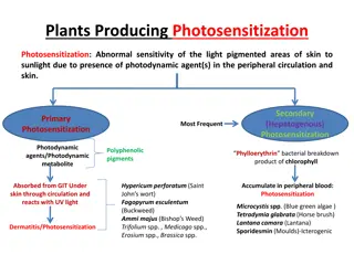
Types of photosensitization
•
Type I: Primary photosensitization
•
Type II: Abnormal porphyrin metabolism associated photosensitization
•
Type III: Hepatogenous photosensitization
Type I: Primary Photosensitization
•
Causes
–
Plants containing helianthrones (e.g. hypericine
in
Hypericum perforatum
; fagopyrin in
Fagopyrum
esculentum
) and furocoumarin pigments (e.g.
Cymopterus
watsonii
and
Ammi majus
), tetracyclines and sulphonamides
•
Examples
–
Phytotoxins from furocoumarin plants exposed to fungi or
other injury may be absorbed into skin which reacts with
UV light
–
Phenothiazine is converted into photoreactive compound
when bypasses the liver, reaches the skin causing
photodermatitis on exposure to sunlight
Type II: Abnormal porphyrin metabolism associated
photosensitization
•
Due to inherited enzyme deficiency, abnormal porphyrin
photodynamic metabolic products like uroporphyrin and
protoporphyrin accumulate in blood and tissues.
•
The uroporphyrin also causes discolouration of bone known as
“osteohaemochromatosis” and teeth called “pink teeth”.
•
Examples
–
Bovine congenital porphyria
–
Bovine haematopoetic protoporphyria
Type III: Hepatogenous photosensitization
•
Hepatogenous photosensitization is caused by impaired
hepatic capacity to excrete phylloerythrin derived from
chlorophyll degradation in the alimentary tract, mainly
affecting herbivores.
•
Causes
–
Hepatocellular damage or injury (Toxic hepatitis due
to
Lantana camara
,
Tribulus terrestris
, plants producing
pyrrolizidine alkaloids, sporidesmins)
–
Inherited hepatic defects
–
Biliary obstruction
–
Infection: Leptospirosis
–
Chemicals: CCl
4
poisoning
•
Gross pathology,
hairless, non-pigmented skin exposed to sun
light shows erythema, edema, blisters, exudation, necrosis and
sloughing of necrotic tissue.
•
Histopathology,
coagulative necrosis of epidermis,
subepidermal vesiculation, swelling of endothelial cells,
fibrinoid degeneration and thrombosis of blood vessels leading
to edema.
•
Secondary bacterial infection culminate in sloughing of
epidermis and adnexae.
Photosensitization in animals is the activation of photodynamic chemicals on the skin by UV or visible light, leading to cellular damage and inflammation. Factors necessary for photosensitization include oxygen, sunlight, specific chemicals, and skin lacking pigments. Types of photosensitization include primary, abnormal porphyrin metabolism-associated, and hepatogenous. Each type has different causes and examples, such as plant toxins, enzyme deficiencies, and liver impairments. Recognizing and understanding the types and causes of photosensitization in animals is crucial for effective management and prevention of this condition.

Download Presentation

Please find below an Image/Link to download the presentation.
The content on the website is provided AS IS for your information and personal use only. It may not be sold, licensed, or shared on other websites without obtaining consent from the author.If you encounter any issues during the download, it is possible that the publisher has removed the file from their server.
You are allowed to download the files provided on this website for personal or commercial use, subject to the condition that they are used lawfully. All files are the property of their respective owners.
The content on the website is provided AS IS for your information and personal use only. It may not be sold, licensed, or shared on other websites without obtaining consent from the author.
E N D
Presentation Transcript
PHOTOSENSITIZATION DR. SANJIV KUMAR ASSTT. PROFESSOR, DEPTT. OF PATHOLOGY, BVC, PATNA
INTRODUCTION Is activation of photodynamic chemicals on the skin by long wave length UV or occasionally by visible light. Necrosis and edema are produced in the exposed areas of skin of animals. The cellular damage by photosensitization is due to release of reactive oxygen species leading to mast cell degranulation and production of chemical mediators of inflammation.
Factors necessary for photosensitization in animals Oxygen Sunlight Photodynamic chemicals Skin devoid of hair or wool and lacking pigments
Types of photosensitization Type I: Primary photosensitization Type II: Abnormal porphyrin metabolism associated photosensitization Type III: Hepatogenous photosensitization
Type I: Primary Photosensitization Causes Plants in esculentum) and furocoumarin pigments (e.g. Cymopterus watsoniiand Ammi majus), tetracyclines and sulphonamides Examples Phytotoxins from furocoumarin plants exposed to fungi or other injury may be absorbed into skin which reacts with UV light Phenothiazine is converted into photoreactive compound when bypasses the liver, reaches the skin causing photodermatitis on exposure to sunlight containing helianthrones perforatum; (e.g. hypericine Fagopyrum fagopyrin in Hypericum
Type II: Abnormal porphyrin metabolism associated photosensitization Due to inherited enzyme deficiency, abnormal porphyrin photodynamic metabolic products like uroporphyrin and protoporphyrin accumulate in blood and tissues. The uroporphyrin also causes discolouration of bone known as osteohaemochromatosis and teeth called pink teeth . Examples Bovine congenital porphyria Bovine haematopoetic protoporphyria
Type III: Hepatogenous photosensitization Hepatogenous photosensitization is caused by impaired hepatic capacity to excrete phylloerythrin derived from chlorophyll degradation in the alimentary tract, mainly affecting herbivores. Causes Hepatocellular damage or injury (Toxic hepatitis due to Lantana camara, Tribulus terrestris, plants producing pyrrolizidine alkaloids, sporidesmins) Inherited hepatic defects Biliary obstruction Infection: Leptospirosis Chemicals: CCl4poisoning
Gross pathology, hairless, non-pigmented skin exposed to sun light shows erythema, edema, blisters, exudation, necrosis and sloughing of necrotic tissue. Histopathology, coagulative necrosis of epidermis, subepidermal vesiculation, swelling of endothelial cells, fibrinoid degeneration and thrombosis of blood vessels leading to edema. Secondary bacterial infection culminate in sloughing of epidermis and adnexae.