Pediatric Intestinal Obstruction: Causes, Symptoms, and Management
Pediatric intestinal obstruction in newborns and older children can result from various conditions such as foregut obstruction, midgut obstruction, and hindgut obstruction. Common causes include esophageal atresia, pyloric stenosis, and Hirschsprung disease. Diagnosis involves clinical evaluation, imaging studies like CXR and ultrasound, and management may require surgical interventions like TOF repair or pyloromyotomy.

Download Presentation

Please find below an Image/Link to download the presentation.
The content on the website is provided AS IS for your information and personal use only. It may not be sold, licensed, or shared on other websites without obtaining consent from the author.If you encounter any issues during the download, it is possible that the publisher has removed the file from their server.
You are allowed to download the files provided on this website for personal or commercial use, subject to the condition that they are used lawfully. All files are the property of their respective owners.
The content on the website is provided AS IS for your information and personal use only. It may not be sold, licensed, or shared on other websites without obtaining consent from the author.
E N D
Presentation Transcript
Ahmed alsghaier Mohammed alsharani Abdulelah Khdr Supervised by : Dr. AymanAlJazaeri
Intestinal obstruction in the newborn infant and older child may be due to a variety of condition: Foregut obstruction Esophageal atresia Pyloric stenosis Malrotation Duodenal atresia Annual pancrease Midgut obstruction : Intestinal atresia Meconiumileus Hindgut obstruction : Hirschsprung Imperforated anus Meconiumplug
Incidence: 1: 5000 live births, 50% associated with anomalies Types: VACTERL Syndrome Symptoms and Signs: Excessive salivation Respiratory Distress Inability to pass NG tube Choking and coughing on feeding
Diagnosis Clinical & CXR Management: Resuscitation Common type Right thoracotomy Division and repair of TOF Primary anastomosis Pure TOF Division and repair Isolated atresia >3 vertebra Staged surgery (gastrostomyand followed in 3-6 months by delayed repair. If fails then need esophageal replacement (stomach or colon)
Risk factors : Family Hx, firstborn male. Age: 2 weeks to 2 months. Sign and symptoms : projectile vomiting , constant hunger, hypochloremic alkalosis,wt loss or poor wt gain. Diagnosis and Treatment : Gastric perstalsis, pyloric mass (olive). Ultrasound. IV line , pyloromytomy.
If the small bowel mesentery is not fixed at the duodenojejunal flexure or ileocecal region Predisposing to valvulus. Ladd s bands may cross the duodenum obstruction. Age : 1/3 (1 week), ( 1 month), 90% ( 1 year ). Present with bilious vomiting. Diagnosis : Upper GI contrast study showing cut off in duodenum; BE showing abnormal postion of cecum .
Maintain patients on nothing by mouth (NPO). Correct fluid and electrolyte deficits. Administer broad-spectrum antibiotics. If a patient has signs of shock, administer appropriate fluids. Surgery : The Ladd procedure: 1-counterclockwise reduction of midgut. 2-splitting of ladds bands. 3-division of peritoneal attachments to cecum. 4-appendectomy.
Divided into: Complete (atresia) Partial ( stenosis) Antenatal diagnosis: Polyhydramnios Dilated stomach and 1st part Duodenum Down syndrome 30%. Symptoms and Signs: vomiting, bilious 80% High gastric aspiration: >30ml
X-rays: Double bobble shadow ( one air in the stomach and other in duodenum). Management: Exclude the Volvulus and resuscitation NGT, Vitamin K, stabilized before surgery Duodeno-duodenostomy
is a malformation where there is a narrowing or absence of a portion of the intestine. This defect can either occur in the small or large intestine. The different types of intestinal atresia are named after their location: Duodenal atresia Jejunal atresia Ilealatresia Colon atresia Duodenal atresia has a strong association with Down syndrome. It is the most common type, followed by ileal atresia.
Causes: The most common cause of non-duodenal intestinal atresia is a vascular accident in utero In the case that the superior mesenteric artery, or another major intestinal artery, is occluded, large segments of bowel can be entirely underdeveloped Dignosis: Intestinal atresiasare often discovered before birth: either during a routine sonogram which shows a dilated intestinal segment due to the blockage, or by the development of polyhydramnios. Treatment laparotomy. If the area affected is small, the surgeon may be able to remove the damaged portion and join the intestine back together. In instances where the narrowing is longer, or the area is damaged and cannot be used for period of time, a temporary Stoma may be placed.
Intestinal obstruction from solid meconium. Bilious vomiting, abd. Distention,failureto pass meconium,Neuhauser s sign. Diagnosis : Family Hx of Cf, AXR, BE. Tratment: Gastrografin enema . If enema is unsuccessful, then enterotomy.
Due to congenital absence of ganglion cells in the distal bowel. Incidence: 1/4500-5000 live births Sex: 4:1 male predominance, Age: 96% Full term & 4% premature Site: Commonly: rectum/rectosigmoid Less commonly: total colonic with or without small intestine
Neonatal: Delayed or failure to pass meconium with low intestinal obstruction. late presentation: Failure to thrive, Poor feeding bloody Diarrhea with abdominal distension and occasionally with enterocolitis. Examination: Abdominal Distension, PR: tight sphincter with gush of loose stool Malnutritedchild, Enterocolitis.
Treatment : Pull-through preceduore Ostomy surgery
imperforate anus or anal atresia is a birth defect in which the rectum is malformed. Its cause is unknown. Diagnosis : p/E X-ray Ultrasound These tests can show your baby's doctor some details, such as where the rectum ends and whether it connects to another structure.
Imperforate anus usually requires immediate surgery to open a passage for feces. Depending on the severity of the imperforate, it is treated either with a perinealanoplastyor with a colostomy.
Pathophysiology. In preschool children. The Dx is more difficult. Fecoliths are more common and can be seen on plain films. Perforation may be rapid as the omentum is less well developed.
They present with : Anorexia, vomiting, abdominal pain(initially central but then localized to the right iliac fossa). DDx: 1. Gastroentritis 2. Mesentric lympadenitis 3. Ovarian pathology 4. UTI 5. Renal stone.
> 3 years, diagnosis is mainly clinical Hx, P/E and CBC+diff < 3 years esp. Infant, difficult Dx Early rupture = (elderly group) Sepsis (fever, WBC) Vomiting (ileus or abscess)
Not needed if the clinical picture is clear. Mainly used in difficult Dx Abdominal XR U/S CT scan
If H&P is doesnt suggest AP Low probability observation + re-evaluation Observation NPO, No analgesia, repeat (Exam + CBC) If AP it will become clear (worse inflammation) Higher probability Laparoscopy or open appendicectomy 5-10% can be normal When normal Look for other ddx Do appendicectomy(even if it s normal)
Late presentation (ruptured) Contained abscess Percutaneousdrain + antibiotics > 6 wks if no abscess appendicectomy Diffuse peritonitis Laparotomyor laparoscopy Abdominal washout Appendicectomy
Resuscitation NPO, NGT IVF IV medication Pain medication Appendectomy. Abscess >> percutaneousdrainage + iv antibiotic.
pathophyisology: Invagenation of proximal bowel into a distal segment. Most common site ileocecalvalve. Presentation with : paroxysmal, sever colicky pain. Sausage shaped mass. Recurrent jelly stool comprising blood stained mucus. Abdominal distention.
Click here to view image full size Dx Best by U/S Target sign, Donut sign. 95% accurate Contrast Enema Dxand treatment Rx Pressure reduction Barium Water Air is most common (less complications) Failed pressure reduction Only few patients (15%) Next is surgical reduction if can t resection Likely PLP
Is a result of the failed obliteration of the omphalomesentericduct Obliteration normally occurs during the 5thweek of embryologic development Can present with obstruction, inflammation, or hemorrhage
More frequent in children (62% <2 yrs) Mostly in males (2/1) Prevalent in 2% of the population Normally occur within 2 feet of ileocecalvalve (though reports of diverticulaup to 180 cm have occurred) Most are approximately 2 inches long
Intestinal hemorrhage, typically painless bleeding. Intestinal obstruction (more common in adults) can be from intussusception, inflammation, adhesions Diverticulitis Perforation Neoplasm (usually sarcoma, carcinoid, adenoca)
50% of MD contain ectopic tissue: Gastric mucosa most common (60-85%) Pancreatic tissue seen in 5-16% Other tissues reported, but rare (colonic, duodenal, jejunal, hepatic, and endometrial) Acidic secretions of gastric tissue or alkaline secretions of pancreatic tissue can cause ulcerations and subsequent bleeding
Technetium pertechnetate(99mTcO4) imaging is the best non-invasive method to diagnose when heterotopicgastric mucosa (HGM) is present AP during OR for AP AP is normal look for Meckle's if found remove.
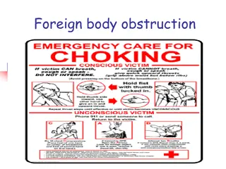
Aspiration and ingestion greatest in children aged 6 months to 4 years concomitant psychiatric problems mental disturbances Younger children may be "fed" foreign bodies by older children Food particle
plain radiograph Most foreign body are radiolucent CT scan or MRI is rarely indicated but may enhance the detection of foreign bodies or complications Bronchoscopy in inhalation If the history and physical findings are typical, no workup is needed
Most swallowed foreign bodies harmlessly pass through the GI tract Surgical therapy for an airway foreign body involves endoscopic removal, usually with a rigid bronchoscope.
Torsion of testis and appendage Infection: epididymitis, epididymo-orchitis, orchitis Trauma Hernia Idiopathic scrotal edema
Torsion occurs when an abnormally mobile testis twists on the spermatic cord, obstructing its blood supply. Patients present with acute onset of severe testicular pain. The ischemia can lead to testicular necrosis if not corrected within 5-6 hours of the onset of pain. Torsion can be intermittent and can undergo spontaneous detorsion. Types: Intravaginal most common, peak incidence b/w 13-16 years of life. Extravaginal- less common and confined to perinatal period.
Color Doppler Complete absence of intratesticular blood flow and normal extratesticular blood flow on color Doppler images is diagnostic High-resolution ultrasonography Nuclear scanning Radioisotope scanning has been reported to be highly accurate for diagnosis of testicular torsion.
in some cases of testicular torsion, manually untwisting the spermatic cord may allow re- establishment of vascular flow. The technique involves manipulating the involved testis so that the anterior surface rotates from medial to lateral. This is termed the "open book" method because the motion resembles opening the cover of a book (for a right testis)
The goals of surgical exploration include (1) confirmation of the diagnosis of torsion (2) detorsion of the involved testis (3) assessment of the viability of the involved testis (4) removal (if nonviable) or fixation (if viable) of the involved testis (5) fixation of the contralateral testis
Testicular atrophy Torsion recurrence Wound infection Subfertility
Visible swelling or bulge in inguinoscrotal aerea May or may not painful After crying or straining Resolve during baby sleep
risk of inguinal hernia: Prematurity and low birth weight (Incidence approaches 50%.) Urologic conditions Hypospadias Epispadias Exstrophyof the bladder Abdominal wall defects Family history Meconiumperitonitis Cystic fibrosis Connective tissue disease Mucopolysaccharidosis Congenital dislocation of the hip
incarceration indicate the following risk patterns: 1. Incarceration occurs in 17% of right-sided hernias and 7% of left-sided hernias. 2. More than 50% of cases of incarceration occur within the first 6 months of life; the risk gradually decreases after age 1 year. 3. Premature infants have twice the risk of incarceration than the general pediatric population. 4. More than two thirds of all incarcerations occur in children younger than 1 year. 5. Girls are more likely to develop incarceration of an inguinal hernia; the incidence in girls is 17.2%, whereas the incidence in boys is 12%.
Ultrasonography: Some advocate the use of ultrasonographyto differentiate between a hydroceleand an inguinal hernia laparoscopy
Parents may be instructed on the application of gentle pressure on the bulge of an inguinal hernia to prevent incarceration until the elective operative repair is performed. (1) high ligation and excision of the patent sac with anatomic closure (2) high ligation of the sac with plication of the floor of the inguinal canal (the transversalisfascia) (3) high ligation of the sac combined with reconstruction of the floor of the canal.
Etiology remains unclear crypts of Morgagni=small infection, or cryptitis abnormal crypts, which predispose to cryptitisand abscess formation Presented with 1-abscess 2-fistula
healthy babies with perianalabscess or fistula require no laboratory studies. Blood counts and cultures should be obtained in all patents who are immunocompromised by any cause Colonoscopy with biopsy may be needed to confirm Crohn disease


.")


")


")


")