Pediatric Cardiovascular Exam Overview
Explore the differences between pediatric and adult cardiovascular exams, highlighting key differences in disease patterns, examination approaches, and objectives. Learn about the importance of distinguishing between routine well-baby exams and evaluations of ill children, along with step-by-step guidance on conducting a pediatric cardiovascular exam.
Download Presentation

Please find below an Image/Link to download the presentation.
The content on the website is provided AS IS for your information and personal use only. It may not be sold, licensed, or shared on other websites without obtaining consent from the author.If you encounter any issues during the download, it is possible that the publisher has removed the file from their server.
You are allowed to download the files provided on this website for personal or commercial use, subject to the condition that they are used lawfully. All files are the property of their respective owners.
The content on the website is provided AS IS for your information and personal use only. It may not be sold, licensed, or shared on other websites without obtaining consent from the author.
E N D
Presentation Transcript
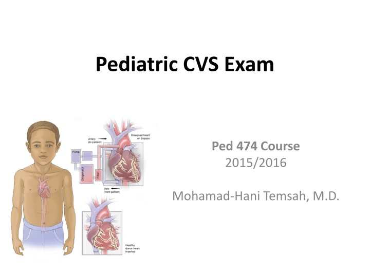
Pediatric CVS Exam Ped 474 Course 2015/2016 Mohamad-Hani Temsah, M.D.
Objectives: To Highlight differences in Pediatric CVS Exam versus Adults To outline the P/E (Principals, Video, Manikin) in order to facilitate application in real patients during your Clinical Teachings To share useful Hints!!
Introduction Main differences between Pediatric & Adult CVS diseases:
Children are not just small adults Some principles of examining children are similar to adult, but there are important differences! Pattern of disease, the approach to the examination and content of the examination are quite different in children. Examination changes as children develop and get older! Sinus (respiratory) arrhythmia is much more prominent. In child, heart occupies relatively more space in the thorax. <7 years: apex beat is in 4th intercostal space to the L of midclavicular line.
Aims / purposes of pediatric examination? It is important to distinguish between: The routine examination of well babies (to screen largely for abnormalities of growth and development). The examination of ill babies (to establish the nature and cause and extent of any illness or injury). The examination of children for other specific purposes such as, for example: To establish fitness for education or certain activities To examine for signs of disease
Step by Step 1. INTRODUCE SELF:
GENERAL INSPECTION Position patient: lying Well or unwell Growth parameters Dysmorphic syndromes/features Scars Chest asymmetry Respiratory rate
3. UPPER LIMBS Nails Clubbing http://www.youtube.com/watch?v=aRwGxvnKSrA Pulses Blood pressure
4. HEAD AND NECK Jugular venous pressure Eyes Conjunctival pallor (anaemia) Scleral icterus (fragmentation haemolysis with artificial valves) Lips: cyanosis Tongue: cyanosis Teeth: caries
CHEST: Inspect Scars Symmetry Apical pulsation
CHEST: Palpate Apex position (beware dextrocardia) (count down the ribs) Heaves (parasternal, substernal, apical) Thrills (suprasternal, supraclavicular) Palpable pulmonary valve closure (pulmonary hypertension)
CHEST: Auscultate (use diaphragm initially, then bell) Areas: apex, parasternal border, pulmonary, aortic areas; roll to left to accentuate mitral murmurs Heart sounds (intensity, splitting) Added sounds Murmurs (systolic, diastolic or continuous, site of maximal intensity, character, grade) Radiation of murmurs: axilla (mitral), neck (aortic), back (pulmonary, coarctation) Variation of murmurs: sitting, inspiration, expiration Maneuvers (if appropriate): Valsalva, exercise Lung fields: adventitious sounds (LVF)
After this, palpate back for Sacral oedema (RVF) Collaterals (if coarctation suspected; also check deep in axillae)
ABDOMEN Liver: (palpate edge and measure span by percussion) Enlarged (RVF) Pulsatile (tricuspid incompetence) Spleen: Enlarged (SBE)
LOWER LIMBS Inspection Clubbing (if not already done) Splinter haemorrhages (if not done) Palpation Ankle oedema (RVF)
OTHER Urinalysis: blood (SBE) Temperature chart (SBE) Fundoscopy for Roth spots if SBE seems likely
INVESTIGATIONS After presenting a differential diagnosis, may request CXR/ECG or other tests to rule in or rule out the DDx
Examination Tips Can establish rapport while checking cyanosis, dyspnea, cough. Can examine teddy bear / toy first. Best examination method by age: Neonates, very young infants: on examining table Up through preschool: lying sit on mother's lap Adolescent: without family present. Kids are impatient, so a systematic full examination may get difficult. Examine the most pertinent area first. Record RR & HR first, before crying may start!! ENT exam more likely to induce a cry so these go last.
The approach to the examination will be determined by the age, level of development and level of understanding of the child. Inspection and observation are the most important parts of the examination. Observations can be made whilst taking the history and establishing rapport. Avoid waking sleeping children. Approach the child at their level, if necessary kneel on the floor.
Start examining peripherally with hands and feet as this is less threatening. Make the examination fun to help with their anxiety. Make sure the child is comfortable, and that your hands, stethoscope and other instruments are warm. Ask parents to assist with dressing or undressing children and be aware of sensitivities about this. Wherever possible avoid unpleasant procedures (for example, rectal examination). These are seldom necessary and can put children off being examined for a lifetime!
The cardiovascular system It may be a good idea to start the physical examination here, if the child is quiet and settled. Letting the child remain seated/held on the parent's or carer's lap, will reassure them: Begin by recording V/S, pulse rate, rhythm, strength and character. Inspect then Palpate the anterior chest wall: apex beat site. Any thrills or heaves? Listen to the 1st heart sound, then the 2nd heart sound, then the sounds between these and then any murmurs between heart sounds. Note the timing, character, loudness, site and distribution of any murmur. Check if this is transmitted to the neck.
Putting Things Together: https://www.youtube.com/watch?v=DvzPh- 2BwrQ&list=PLPHYOKOlW_qwpRAbMunOlXhr v-3_kzQm1 https://www.youtube.com/watch?v=eBnzjerIHj0
Ref. Examination Paediatrics: A Guide to Paediatric Training (Second Edition) http://www.patient.co.uk/doctor/Paediatric- Examination.htm http://www.clinicalexam.com/pda/peds_exam.htm
Best Wishes in Ped 474, and in YOUR Careers! Hani mtemsah@ksu.edu.sa
