Parts of the Brain and Their Functions


Part I: Parts of the Brain
and How they Work
•
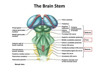
The Brain Stem
– This looks like a stalk rising out
of the spinal cord.
•
Pathways to and from upper areas include the
medulla
and
pons
.
•
Pons
– involve sleeping, waking, and dreaming.
•
Medulla
– responsible for bodily functions that do
not have to be consciously willed, such as breathing
and heart rate.
•
RAS – The reticular activating system extends
upward from the core of the brain. These neurons
extend to the center of the brain. Here when
something happens it demands the attention of the
higher areas, without them we would not be alert or
conscious.

Parts of the Brain
•
The Cerebellum –
Also known as the “lesser
brain,” this gives us balance and coordinates the
muscles so that movement is smooth and precise.
•
If your cerebellum were damaged you would
probably be clumsy and uncoordinated.
•
This part of the brain also is involved in
remembering simple skills and acquired reflexes
(tying your shoes, juggling a soccer ball).
•
In addition, it also plays a part in complex tasks and
analyzing sensory information.
Parts of the Brain
•
The Thalamus –
Known as the traffic officer of
the brain, the Thalamus directs sensory
messages that come into the brain to high
centers.
•
For example – A sunset would send signals
directed to vision area, sound signals sends
information to the auditory area.
•
The only sense that completely bypasses the
thalamus is the sense of smell, which has its own
private switching station, the
olfactory bulb
.
Parts of the Brain
•
They Hypothalamus and the Pituitary Gland –
“Hypo” means under. This is involved in drives
associated with survival of the individual and species.
•
It regulates body temperature by triggering sweating or
shivering, and controls the operations of the atomic
nervous system.
•
It also contains the biological clock that controls the
body’s daily rhythms.
•
Hanging down from the hypothalamus is the
pituitary
gland
.
•
Also known as the “master gland” because the hormones
it secretes effect other endocrine glands by sending
hormonal messages out to these glands.
•
TOGETHER THESE MAKE UP THE LIMBIC SYSTEM.
Parts of the Brain
•
The Amygdala –
(Ancient word for almond) is
responsible for evaluating sensory information,
quickly determining its emotional importance,
and contributing to the initial decision to
approach or withdraw from a person or
situation.
•
Instantly assesses danger or a threat, mediates
anxiety or depression. PET scans find that
depressed and anxious patients show increased
neural activity in the limbic structure.
Parts of the Brain
•
The Hippocampus –
(Means seahorse in Latin, named
after its shape).
•
This structure compares sensory information with what the
brain has learned to expect about the world.
•
When expectations are met, there is no need for neural alarm
every time something happens around you.
•
It has also been called the “gateway to memory.” The
Hippocampus enables us to form spatial memories so that we
can accurately navigate through our environment.
•
In addition, it also forms new memories to help you identify
and create new memories and facts.
•
This information is stored in the cerebral cortex. For
examples, when you recall information you remember things
like tone, appearance, and location.
Parts of the Brain
•
The Cerebrum –
Where the higher forms of
thinking take place.
•
Divided into two separate halves, or
cerebral
hemispheres
, connected by a large band of fibers
called the
corpus callosum
.
•
In general, the right hemisphere is in charge of
the left side of the body and the left hemisphere
is in charge of the right side of the body.
•
The two hemispheres also have somewhat
different tasks.
Part II: Lobes of the Cerebrum
•
The Cerebral Cortex -
A collection of several thin
layers of cells covering the cerebrum; it is largely
responsible for higher mental functions.
•
Contains both grey and white matter, referring to
the types of tissue located here. Some are more grey
and some are more white.
•
It is only about 3 millimeters thick, yet it contains
three-fourths of all the cells in the human brain.
•
The Cortex
has many crevasses and wrinkles, which
enable it to contain its billions of neurons without
requiring us to have the heads of giants - heads that
would be too big to allow us to be born.
Lobes of the Cortex
•
Occipital Lobes –
are at the lower back part of the brain. They
contain the
visual cortex
,
where visual signals are processed
.
Damaged to the visual cortex can cause impaired visual recognition
or even blindness.
•
Parietal Lobes –
They contain the
somatosensory cortex
,
which
receives information about pressure, pain, touch and temperature
from all over the body.
Parts are also involved in attention and
various mental operations.
•
Temporal Lobes –
are at the sides of the brain, just abbbove the
ears, behind the temples. They are involved in memory, perception,
and emotion, and they contain the autidory cortex which processes
sounds.
•
An area of the left
temporal lobe
known as
Wernicke’s area
is
involved in language comprehension.
•
Frontal Lobes –
Located toward the front of the brain just under
the skull in the area of the forehead. They contain the
motor cortex
,
which issues orders to the 600 muscles in the body that produce
voluntary movement.
In the
left frontal
lobe, a region known as
Broca’s area
handles speech production.
During short-term memory
tasks, areas in the frontal lobes are especially active. The frontal
lobes are also involved in emotion and the ability to make plans =
think creatively, and take initiative.
The brain consists of different parts with unique roles. The Brain Stem controls essential functions like breathing, while the Cerebellum aids in balance and muscle coordination. The Thalamus directs sensory messages, the Hypothalamus regulates survival drives, and the Amygdala evaluates emotional responses. The Hippocampus compares sensory information with learned expectations, shaping our perception of the world.

Download Presentation

Please find below an Image/Link to download the presentation.
The content on the website is provided AS IS for your information and personal use only. It may not be sold, licensed, or shared on other websites without obtaining consent from the author. Download presentation by click this link. If you encounter any issues during the download, it is possible that the publisher has removed the file from their server.
E N D
Presentation Transcript
Part I: Parts of the Brain and How they Work The Brain Stem This looks like a stalk rising out of the spinal cord. Pathways to and from upper areas include the medulla and pons. Pons involve sleeping, waking, and dreaming. Medulla responsible for bodily functions that do not have to be consciously willed, such as breathing and heart rate. RAS The reticular activating system extends upward from the core of the brain. These neurons extend to the center of the brain. Here when something happens it demands the attention of the higher areas, without them we would not be alert or conscious.
Parts of the Brain The Cerebellum Also known as the lesser brain, this gives us balance and coordinates the muscles so that movement is smooth and precise. If your cerebellum were damaged you would probably be clumsy and uncoordinated. This part of the brain also is involved in remembering simple skills and acquired reflexes (tying your shoes, juggling a soccer ball). In addition, it also plays a part in complex tasks and analyzing sensory information.
Parts of the Brain The Thalamus Known as the traffic officer of the brain, the Thalamus directs sensory messages that come into the brain to high centers. For example A sunset would send signals directed to vision area, sound signals sends information to the auditory area. The only sense that completely bypasses the thalamus is the sense of smell, which has its own private switching station, the olfactory bulb.
Parts of the Brain They Hypothalamus and the Pituitary Gland Hypo means under. This is involved in drives associated with survival of the individual and species. It regulates body temperature by triggering sweating or shivering, and controls the operations of the atomic nervous system. It also contains the biological clock that controls the body s daily rhythms. Hanging down from the hypothalamus is the pituitary gland. Also known as the master gland because the hormones it secretes effect other endocrine glands by sending hormonal messages out to these glands. TOGETHER THESE MAKE UP THE LIMBIC SYSTEM.
Parts of the Brain The Amygdala (Ancient word for almond) is responsible for evaluating sensory information, quickly determining its emotional importance, and contributing to the initial decision to approach or withdraw from a person or situation. Instantly assesses danger or a threat, mediates anxiety or depression. PET scans find that depressed and anxious patients show increased neural activity in the limbic structure.
Parts of the Brain The Hippocampus (Means seahorse in Latin, named after its shape). This structure compares sensory information with what the brain has learned to expect about the world. When expectations are met, there is no need for neural alarm every time something happens around you. It has also been called the gateway to memory. The Hippocampus enables us to form spatial memories so that we can accurately navigate through our environment. In addition, it also forms new memories to help you identify and create new memories and facts. This information is stored in the cerebral cortex. For examples, when you recall information you remember things like tone, appearance, and location.
Parts of the Brain The Cerebrum Where the higher forms of thinking take place. Divided into two separate halves, or cerebral hemispheres, connected by a large band of fibers called the corpus callosum. In general, the right hemisphere is in charge of the left side of the body and the left hemisphere is in charge of the right side of the body. The two hemispheres also have somewhat different tasks.
Part II: Lobes of the Cerebrum The Cerebral Cortex - A collection of several thin layers of cells covering the cerebrum; it is largely responsible for higher mental functions. Contains both grey and white matter, referring to the types of tissue located here. Some are more grey and some are more white. It is only about 3 millimeters thick, yet it contains three-fourths of all the cells in the human brain. The Cortex has many crevasses and wrinkles, which enable it to contain its billions of neurons without requiring us to have the heads of giants - heads that would be too big to allow us to be born.
Lobes of the Cortex Occipital Lobes are at the lower back part of the brain. They contain the visual cortex, where visual signals are processed. Damaged to the visual cortex can cause impaired visual recognition or even blindness. Parietal Lobes They contain the somatosensory cortex, which receives information about pressure, pain, touch and temperature from all over the body. Parts are also involved in attention and various mental operations. Temporal Lobes are at the sides of the brain, just abbbove the ears, behind the temples. They are involved in memory, perception, and emotion, and they contain the autidory cortex which processes sounds. An area of the left temporal lobe known as Wernicke s area is involved in language comprehension. Frontal Lobes Located toward the front of the brain just under the skull in the area of the forehead. They contain the motor cortex, which issues orders to the 600 muscles in the body that produce voluntary movement. In the left frontal lobe, a region known as Broca s area handles speech production. During short-term memory tasks, areas in the frontal lobes are especially active. The frontal lobes are also involved in emotion and the ability to make plans = think creatively, and take initiative.