Optimizing ECG Quality: Patient Preparation & Lead Placement
Learn the importance of preparing the patient for ECG to enhance signal quality, including explaining the procedure, exposing the chest, removing interfering objects, shaving chest hair if necessary, and skin preparation for electrode placement. Discover lead placement techniques and anatomical landmarks for accurate ECG readings.

Download Presentation

Please find below an Image/Link to download the presentation.
The content on the website is provided AS IS for your information and personal use only. It may not be sold, licensed, or shared on other websites without obtaining consent from the author. If you encounter any issues during the download, it is possible that the publisher has removed the file from their server.
You are allowed to download the files provided on this website for personal or commercial use, subject to the condition that they are used lawfully. All files are the property of their respective owners.
The content on the website is provided AS IS for your information and personal use only. It may not be sold, licensed, or shared on other websites without obtaining consent from the author.
E N D
Presentation Transcript
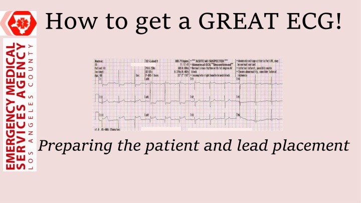
How to get a GREAT ECG! Preparing the patient and lead placement
Objectives Review patient preparation techniques for optimizing ECG quality Define anatomical landmarks for ECG lead placement Demonstrate correct ECG lead placement
Preparing the Patient 1.Make the patient as relaxed and comfortable as possible 2.Explain the procedure *relaxed patient = better ECG quality
Preparing the Patient 4.Expose the chest 5.Remove garments 6. Remove accessories that may interfere with ECG
Preparing the Patient Remove hair Shave interfering chest hair, if applicable Removing excess hair allows electrode gel to penetrate the skin resulting in a stronger signal
Preparing the Patient 1. Wipe down skin with single use washcloth or gauze 2. Vigorously wipe skin prior to electrode placement -Reduces skin oil -Abrades the top skin layers for better contact
Lead Placement Position The Patient - Place patient supine (or closest position tolerated) for optimal lead placement
Lead Placement Precordial Leads
Lead Placement First Step: Locate the Angle of Louis -Find your jugular notch (valley at base of throat) -Move finger down until you feel a ridge (that s it!)
Lead Placement Found your angle? Great! It s time to start applying the leads! Tips before we begin: -Minimize the time the electrode leads are exposed to air -In patients with breast/chest tissue, do NOT alter the positioning of the electrodes. Place the leads on the breast tissue if lead positioning is compromised by going below.
Lead Placement Second Step: Find V1 1. Move from the Angle of Louis to the gap on the right **this is your 2nd intercostal space** 2. Move down two rib spaces to the 4th intercostal space 3. Place your lead where the space meets the right sternal border
Lead Placement Third Step: Place Leads V2-V4 1. V2: Place next to sternum in the 4th intercostal space on LEFT side 2. V4: Place in 5th intercostal space on the left, at the mid-clavicular line 3. V3: Place mid-way between V2 and V4
Lead Placement Fourth Step: Place Leads V5 and V6 1. V5: Place next to v4 in the same horizontal plane at the left anterior axillary line (in-line with crease of arm) 2. V6: Place next to v5 in the same horizontal plane at the left mid-axillary line (in the center of the under-arm area)
Lead Placement The Final step: Limb Leads They must be placed ON THE LIMBS, not the torso
Lead Placement The Final step: Limb Leads - Place as proximal as possible - Upper extremity leads should be placed distal to the deltoid - Lower extremity leads should be placed distal to the inguinal line
Acquiring the ECG Prepare For Excellence! Wire Management - Ensure the wires are fanned out - Ensure the wires are slack and free of obstruction - Connect lead clips to clothing to minimize movement Position Patient - Supine or semi-recumbent if tolerated - Head back on stretcher - Arms resting at sides
Acquiring a GREAT ECG You re almost ready! -Ensure the patient is relaxed -Ensure the patient is not talking or moving -Ensure that YOU are not moving (or driving) Capture a GREAT ECG!
Types of Artifact Motion artifact Muscle artifact Missing lead Electromagnetic interference
Motion Artifact Low Frequency Isolated possibly due to patient motion, try to get patient to stay still Prolonged - possibly due to respiration (have patient hold their breath) High Frequency Possibly due to ambulance motion, are you transporting? If not, is there something causing the patient to move rapidly?
Muscle Artifact Muscle Tension Determine and treat underlying cause: i.e. Is patient in pain? Are they anxious? Muscle Tremor Is the patient shivering? Consider treating the cause. Electrode on a spasming muscle? Consider moving it if possible
Missing Lead Check for: -Leads and wires connected -Dry electrodes? (poor contact) -Cable failure? (consider swapping for spare) -Connectivity issue? (chest hairy, skin oily/wet?) -Machine issue? (consider service call)
Electromagnetic Interference Interference such as this may be seen next to a power line Interference such as this may be seen next to a cell phone or other such devices
So, you got a great ECG, now what? Make sure the patient s age and gender are entered correctly as this can affect software interpretation. Review the ECG yourself; what is your interpretation? If you cannot interpret the ECG due to quality issues, neither can the software REPEAT! Keep the leads on! Be prepared to repeat the ECG, especially if you have a high suspicion for STEMI, an initial non-diagnostic ECG, or if the patient s clinical condition changes.
Summary Prepare patient by helping them relax Prep patient s skin for electrode placement Proper lead placement is essential: Precordial lead landmarks should be palpated Limb leads must be placed on the extremities, NOT the torso Reduce patient movement and activity Recognize and know how to troubleshoot artifact Be prepared to repeat the ECG, especially if paramedic s suspicion for STEMI is high


