Mycobacterium: Causes and Impact of Tuberculosis

Mycobacterium
assist. Prof.
Zainab Abdul jabar
Aldhaher
According to the World Health Organization (WHO),
nearly 2 billion people,
one-third of the world
’
s
population
, have disease caused by mycobacteria,
particularly tuberculosis.
Mycobacteria are widespread both in the environment
and in animals and cause two major human diseases –
tuberculosis and leprosy. They are aerobic, acid-fast
bacilli (not stained by the Gram stain because of the high
lipid component of the cell wall). The major medically
important pathogens are:
•
Mycobacterium tuberculosis
, the agent of
tuberculosis
;
one of the top three infectious diseases affecting humans
globally
Pathogenicity
This organism is the agent of tuberculosis, a
chronic, granulomatous, slowly progressive
infection, usually of the lungs; eventually, many
other organs and tissues may be affected.
A pandemic disease, tuberculosis is especially
common in the developing world owing to HIV
infection (15–20% of individuals with HIV disease
may have tuberculosis).
Mycobacterium
•
Mycobacterium
is a
genus
of
Actinobacteria
, given
its own family, the Mycobacteriaceae.
•
The genus includes
pathogens
known to cause
serious diseases in mammals, including:
•
tuberculosis
(
Mycobacterium tuberculosis
)
•
leprosy
(
Mycobacterium leprae
).
•
mycobacterium tuberculosis
(MTB) is a
pathogenic
bacterial
species in the genus
Mycobacterium
and the causative agent of
most cases of
tuberculosis
(TB)First
discovered in 1882 by
Robert Koch
,
•
M. tuberculosis
has an unusual, waxy coating on its
cell surface (primarily
mycolic acid
), which makes
the cells impervious to
Gram staining
, so
acid-fast
detection techniques are used, instead. The
physiology of
M. tuberculosis
is highly
aerobic
and
requires high levels of oxygen.
Microbiologic characteristics
•
Mycobacteria are
aerobic
and nonmotile
bacteria.
•
Mycobacteria do not contain
endospores
or
capsules
and are usually considered
Gram-
positive
.
•
All
Mycobacterium
species share a characteristic
cell wall
, thicker than in many other bacteria,
which is
hydrophobic
, waxy, and rich in
mycolic
acids
/mycolates.
•
Many
Mycobacterium
species adapt readily to
growth on very simple
substrates
, using
ammonia
or
amino acids
as nitrogen sources and
glycerol
as
a carbon source in the presence of mineral salts.
Optimum growth temperatures vary widely
according to the species and range from 25 °C to
over 50 °C.
•
Some species can be very difficult to
culture
(i.e.
they are
fastidious
),
•
M. tuberculosis
requires oxygen to grow
. It does
not retain any bacteriological stain due to high
lipid content in its wall, and thus is neither
Gram-positive nor Gram-negative; hence
Ziehl-
Neelsen staining
, or acid-fast staining,
•
M. tuberculosis
divides every 15–20 hours, which
is extremely slow compared to other bacteria,
which tend to have division times measured in
minutes (
Escherichia coli
can divide roughly every
20 minutes). It is a small
bacillus
that can
withstand weak
disinfectants
and can survive in a
dry state for weeks. Its unusual cell wall, rich in
lipids
(e.g.,
mycolic acid
), is likely responsible for
this resistance and is a key virulence factor.
symptoms
•
Fever
•
Malaise – tired, achy
•
Lung degeneration – pneumonia
•
Chronic cough
•
Septicemia, multiple organ failure
•
Weight loss despite increased appetite –
“consumption”
Virulence Factors
•
Waxy cell wall.
•
Major factor is ability to invade and survive
within macrophages as surface protein called
“exported repetitive protein” prevents
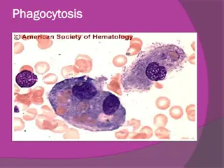
phagosome from joining with lysosome.
•
It produces no exotoxins or no LPS.
Pathogenicity
•
The organism generally transmitted by droplets
from person with active case of tuberculosis.
•
The microorganism is very stable in sputum
droplets and can remain viable in very even dry
sputum for up 6 days.
•
M. tuberculosis in droplets is then inhaled and
reach the highly aerobic environment of the lung
where it produce non specific pneumonitis
.
•
Inflammation occurs such that more
phagocytes travel to the site this is called an
“exudative lesion” end up with a mass of live
and dead bacteria, live and dead phagocytes
surrounded by an outer layer of macrophages
called a granuloma – due to large number of
granulocytes, granuloma becomes surrounded
by fibrin which calcifies called a tubercle, can
be seen by chest X – ray. The infection may
stop at this point and the individual may have
no more symptoms
•
The tubercle can break though the lung into
blood vessels and then be disseminated
throughout the body – becomes systemic
infection, 50% mortality rate.
•
Tubercle can be coughed up and swallowed,
becoming systemic via the gastrointestinal
tract
•
Tubercle can burst years after primery
infection –
Reactivation TB
•
diagnosis
1.
demonstration of acid fast bacilli in smear made
from sputum sample is indicative of tuberculosis.
2.
M.tuberculosis can be culture from suptum or other
contaminated fluids onto egg yolk containing agar or
onto oleic acid albumin agar following 2-4 weeks of
incubation.
•
The
Löwenstein–Jensen medium
, is a growth
medium specially used for culture of
Mycobacterium
species, notably
Mycobacterium tuberculosis
.
Löwenstein–Jensen medium
Löwenstein-Jensen agar
When grown on this medium appears as brown,
granular colonies (sometimes called "buff, rough
and tough"). The medium must be incubated for a
significant length of time, usually four weeks, due
to the slow doubling time of
M. tuberculosis
(15–
20 hours) compared with other bacteria.
3.
Chest X – ray
4.
Tuberculin skin test (Mantoux Test)
Purified protein derivative (PPD), part of cell wall is
injected under the skin, 24 – 48 hours measure the
size of the welt that forms. Cell have been primed
and recognize the PPD
.
o
Positive reaction means you have been
exposed to the organism:
•
Could have active infection
•
Could have been infected but were one of the
90% who are Asymptomatic.
•
You have been immunized
Antigenic structure:
•
The mycobacterial antigens have been classified
as:
•
1- Soluble (cytoplasmic) and insoluble (cell wall
lipid bound).
•
2- Carbohydrates or proteins
•
3- by their distribution within the genus
Treatment
•
Antibiotic sensitivity and control
Long-term therapy (6–9 months) with antituberculous
drugs (isoniazid, rifampicin, pyrazinamide, ethambutol).
As drug resistance is growing and a persistent problem,
combination therapy
should always be given. Tubercle
bacilli resistant to a number of antituberculous drugs
(
multidrug-resistant tuberculosis
(MDR-TB)) is a
growing problem. Hence, regimentation of drug delivery is
a cornerstone of managing the disease, which is achieved
by a global programme termed
directly observed therapy
(
DOT
). Prevention is by
bacille Calmette
–
Gu
é
rin
(
BCG
)
vaccination
containing live attenuated organisms, in
childhood.
vaccine
BCG (Bacilli Calmete Guerin) vacine
•
Made to stimulate the cell mediated immune
response as
M. tuberculosis
is largely an
intracellular pathogen
•
Uses live attenuated
M. bovis
, causes TB in cows
•
Most effective in children, given as part of early
childhood regime in endemic regions
•
Some cases of human contracting
M. bovis
from
vaccine, or from drinking unpasteurized milk from
infected cow.
Mycobacterium is a genus of Actinobacteria known for causing serious diseases in mammals, including tuberculosis (TB) and leprosy. Tuberculosis, caused mainly by Mycobacterium tuberculosis, is a chronic infection that affects the lungs and various organs, especially prominent in developing regions due to its association with HIV. The high lipid content in the cell wall of mycobacteria makes them resistant to Gram staining, requiring alternative detection methods. Despite being aerobic and nonmotile, mycobacteria are significant pathogens affecting billions worldwide.

Download Presentation

Please find below an Image/Link to download the presentation.
The content on the website is provided AS IS for your information and personal use only. It may not be sold, licensed, or shared on other websites without obtaining consent from the author.If you encounter any issues during the download, it is possible that the publisher has removed the file from their server.
You are allowed to download the files provided on this website for personal or commercial use, subject to the condition that they are used lawfully. All files are the property of their respective owners.
The content on the website is provided AS IS for your information and personal use only. It may not be sold, licensed, or shared on other websites without obtaining consent from the author.
E N D
Presentation Transcript
Mycobacterium assist. Prof. Zainab Abdul jabar Aldhaher
According to the World Health Organization (WHO), nearly 2 billion people, one-third of the world s population, have disease caused by mycobacteria, particularly tuberculosis. Mycobacteria are widespread both in the environment and in animals and cause two major human diseases tuberculosis and leprosy. They are aerobic, acid-fast bacilli (not stained by the Gram stain because of the high lipid component of the cell wall). The major medically important pathogens are: Mycobacterium tuberculosis, the agent of tuberculosis; one of the top three infectious diseases affecting humans globally
Pathogenicity This organism is the agent of tuberculosis, a chronic, granulomatous, slowly progressive infection, usually of the lungs; eventually, many other organs and tissues may be affected. A pandemic disease, tuberculosis is especially common in the developing world owing to HIV infection (15 20% of individuals with HIV disease may have tuberculosis).
Mycobacterium Mycobacterium is a genus of Actinobacteria, given its own family, the Mycobacteriaceae. The genus includes pathogens known to cause serious diseases in mammals, including: tuberculosis (Mycobacterium tuberculosis) leprosy (Mycobacterium leprae).
mycobacterium tuberculosis (MTB) is a pathogenic bacterial species in the genus Mycobacterium and the causative agent of most cases of tuberculosis (TB)First discovered in 1882 by Robert Koch,
M. tuberculosis has an unusual, waxy coating on its cell surface (primarily mycolic acid), which makes the cells impervious to Gram staining, so acid-fast detection techniques are used, instead. The physiology of M. tuberculosis is highly aerobic and requires high levels of oxygen.
Microbiologic characteristics Mycobacteria are aerobic and nonmotile bacteria. Mycobacteria do not contain endospores or capsules and are usually considered Gram- positive. All Mycobacterium species share a characteristic cell wall, thicker than in many other bacteria, which is hydrophobic, waxy, and rich in mycolic acids/mycolates.
Many Mycobacterium species adapt readily to growth on very simple substrates, using ammonia or amino acids as nitrogen sources and glycerol as a carbon source in the presence of mineral salts. Optimum growth temperatures vary widely according to the species and range from 25 C to over 50 C. Some species can be very difficult to culture (i.e. they are fastidious),
M. tuberculosis requires oxygen to grow. It does not retain any bacteriological stain due to high lipid content in its wall, and thus is neither Gram-positive nor Gram-negative; hence Ziehl- Neelsen staining, or acid-fast staining,
M. tuberculosis divides every 1520 hours, which is extremely slow compared to other bacteria, which tend to have division times measured in minutes (Escherichia coli can divide roughly every 20 minutes). It is a small bacillus that can withstand weak disinfectants and can survive in a dry state for weeks. Its unusual cell wall, rich in lipids (e.g., mycolic acid), is likely responsible for this resistance and is a key virulence factor.
symptoms Fever Malaise tired, achy Lung degeneration pneumonia Chronic cough Septicemia, multiple organ failure Weight loss despite increased appetite consumption
Virulence Factors Waxy cell wall. Major factor is ability to invade and survive within macrophages as surface protein called exported repetitive protein prevents phagosome from joining with lysosome. It produces no exotoxins or no LPS.
Pathogenicity The organism generally transmitted by droplets from person with active case of tuberculosis. The microorganism is very stable in sputum droplets and can remain viable in very even dry sputum for up 6 days. M. tuberculosis in droplets is then inhaled and reach the highly aerobic environment of the lung where it produce non specific pneumonitis.
Inflammation occurs such that more phagocytes travel to the site this is called an exudative lesion end up with a mass of live and dead bacteria, live and dead phagocytes surrounded by an outer layer of macrophages called a granuloma due to large number of granulocytes, granuloma becomes surrounded by fibrin which calcifies called a tubercle, can be seen by chest X ray. The infection may stop at this point and the individual may have no more symptoms
The tubercle can break though the lung into blood vessels and then be disseminated throughout the body becomes systemic infection, 50% mortality rate. Tubercle can be coughed up and swallowed, becoming systemic via the gastrointestinal tract Tubercle can burst years after primery infection Reactivation TB
diagnosis 1. demonstration of acid fast bacilli in smear made from sputum sample is indicative of tuberculosis. 2. M.tuberculosis can be culture from suptum or other contaminated fluids onto egg yolk containing agar or onto oleic acid albumin agar following 2-4 weeks of incubation. The L wenstein Jensen medium, is a growth medium specially used for culture of Mycobacterium species, notably Mycobacterium tuberculosis.
LwensteinJensen medium L wenstein-Jensen agar
When grown on this medium appears as brown, granular colonies (sometimes called "buff, rough and tough"). The medium must be incubated for a significant length of time, usually four weeks, due to the slow doubling time of M. tuberculosis (15 20 hours) compared with other bacteria. 3. Chest X ray 4. Tuberculin skin test (Mantoux Test) Purified protein derivative (PPD), part of cell wall is injected under the skin, 24 48 hours measure the size of the welt that forms. Cell have been primed and recognize the PPD.
o Positive reaction means you have been exposed to the organism: Could have active infection Could have been infected but were one of the 90% who are Asymptomatic. You have been immunized
Antigenic structure: The mycobacterial antigens have been classified as: 1- Soluble (cytoplasmic) and insoluble (cell wall lipid bound). 2- Carbohydrates or proteins 3- by their distribution within the genus
Treatment Antibiotic sensitivity and control Long-term therapy (6 9 months) with antituberculous drugs (isoniazid, rifampicin, pyrazinamide, ethambutol). As drug resistance is growing and a persistent problem, combination therapy should always be given. Tubercle bacilli resistant to a number of antituberculous drugs (multidrug-resistant tuberculosis (MDR-TB)) is a growing problem. Hence, regimentation of drug delivery is a cornerstone of managing the disease, which is achieved by a global programme termed directly observed therapy (DOT). Prevention is by bacille Calmette Gu rin (BCG) vaccination containing live attenuated organisms, in childhood.
vaccine BCG (Bacilli Calmete Guerin) vacine Made to stimulate the cell mediated immune response as M. tuberculosis is largely an intracellular pathogen Uses live attenuated M. bovis, causes TB in cows Most effective in children, given as part of early childhood regime in endemic regions Some cases of human contracting M. bovis from vaccine, or from drinking unpasteurized milk from infected cow.

,")


 is a")