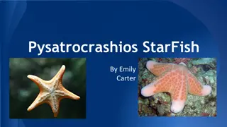
Morphology of Starfish at Mrs. K.S.K. College, Beed
Mrs. K.S.K. College in Beed, under the Department of Zoology, delves into the fascinating morphology of starfish. The external features of starfish, including its shape, size, and color variations, are explored. Specific focus is given to the radially symmetrical and pentamerous body structure of starfish, detailing the central disc and elongated rays or arms. The oral surface of the starfish is examined, highlighting structures such as the mouth, ambulacral grooves, tube feet, and ambulacral spines.
Download Presentation

Please find below an Image/Link to download the presentation.
The content on the website is provided AS IS for your information and personal use only. It may not be sold, licensed, or shared on other websites without obtaining consent from the author.If you encounter any issues during the download, it is possible that the publisher has removed the file from their server.
You are allowed to download the files provided on this website for personal or commercial use, subject to the condition that they are used lawfully. All files are the property of their respective owners.
The content on the website is provided AS IS for your information and personal use only. It may not be sold, licensed, or shared on other websites without obtaining consent from the author.
E N D
Presentation Transcript
Mrs.K.S.K.College,Beed Dept.of Zoology Topic Morphology of star Fish Dr.A.N.Shelke
External Features of star fish Shape, Size and Colour Asterias has a radially symmetrical and pentamerous body. The body consists of a central, pentagonal central disc from which radiate out five elongated, tapering, symmetrical spaced projections, the rays or arms. In some genera, the number of arms may be more than five, for example, there are 7-14 arms in Solaster and more than 40 arms in Heliaster. The size varies from 10-20 cm in diameter though some forms may be much smaller or longer. The colour is variable having shades of yellow, orange, brown and purple. The body has two surfaces, the upper convex and much darker side is called the aboral or abactinal surface.
External Features of star fish Shape, Size and Colour The lower surface is flat, less pigmented and is called the oral or actinal surface. The oral and aboral surfaces are not the ventral and dorsal surfaces but correspond to the left and right sides of the bilaterally symmetrical larva. The axes occupied by the arms are known as radii and the regions of the central disc between the arms are inter-radii. A well defined head is entirely absent.
Oral Surface The oral surface bears the following structures: 1. Mouth: On the oral surface, in the centre of the pentagonal central disc is an aperture, the actinosome or mouth. It is a pentagonal aperture with five angles, each directed towards an arm. The mouth is surrounded by a soft and delicate membrane, the peristomial membrane or peristome and is guarded by five groups of oral spines or mouth papillae. 2. Ambulacral Grooves: From each angle of the mouth radiates a narrow groove called the ambulacral groove which runs all along the middle of oral surface of each arm.
Oral Surface 3. Tube Feet or Podia: Each ambulacral groove contains four rows of locomotory, food capturing, respiratory and sensory organs called tube feet or podia. The tube feet are soft, thin- walled, tubular, retractile structures provided with terminal discs or suckers. The suckers function as suction cups to afford a firm attachment on the surface to which they are applied. 4. Ambulacral Spines: Each ambulacral groove is bordered and guarded laterally by 2 or 3 rows of movable calcareous ambulacral spines which are capable of closing over the groove. Near the mouth, these spines often become larger, stouter, assemble in five groups, one at each inter-radius of disc and are called mouth papilla. Outside the ambulacral spines are three rows of stout immovable spines, beyond which occurs another series of marginal spines along the borders of the arms demarcating the oral from the aboral surface.
Oral Surface 5. Sense Organs: Sense organs include five unpaired terminal tentacles and five unpaired eye spots. The tip of each arm bears a small median, non- retractile and hollow projection, the terminal tentacle. It acts as a tactile and olfactory organ. At the base of each tentacle occurs a bright red photo-sensitive eye spot made up of several ocelli.
Aboral Surface The aboral surface bears following structures: 1. Anus: A minute circular aperture, called the anus, is situated close to the centre of the central disc of aboral surface.
Aboral Surface 2. Madreporite: At the aboral surface of the central disc occurs a flat, sub-circular, asymmetrical and grooved plate called madreporite plate or madreporite between the bases of two of the five arms. The surface of madreporite is marked by a number of radiating, narrow, straight or slightly wavy grooves with pores in them. The madreporite is, thus, a sieve-like porous plate and it leads to the stone canal of water vascular system. The number of madreporite to an individual though remains one, but the presence of more than one madreporite in some species is due to the increase in number of arms beyond the normal number of five. The two arms having madreporite between their bases are collectively referred to as a bivium and the other three arms as a trivium. The symmetrical position of madreporite, thus, converts the radial symmetry of Asterias into bilateral symmetry.
Aboral Surface 3. Spines: The entire aboral surface is covered with numerous short, stout, blunt, calcareous spines or tubercles. The spines are variable in size and are arranged in irregular rows running parallel to the long axes of the arms. The spines are supported by the irregularly-shaped calcareous plates or ossicles which remain buried in the integument and form the endoskeleton
Aboral Surface 4. Papulae or Gills: Between the ossicles of integument are present a large number of minute dermal pores. Through each dermal pore projects out a very small, delicate, tubular or conical, finger-like or thread-like, thin-walled, membranous and retractile projection called the dermal branchia or gill or papula. The papulae are hollow evaginations of the body wall and their lumen remains in continuation with the coelom. They are internally lined by coelom. They have respiratory, as well as excretory functions
Aboral Surface 5. Pedicellariae: Besides the spines and gills, entire aboral surface is covered by many whitish modified spine-like tiny pincers or jaws called pedicellariae. The oral surface also bears pedicellariae. Each pedicellaria consists of a long or short, stout, flexible stalk having no internal calcareous support. The stalk bears three calcareous ossicles or plates a basilar piece or plate at the extremity of the stalk and jaws or valves which remain movably articulated with the basilar piece and serrated along their apposed edges. Pedicellariae having three calcareous pieces and a stalk are called forcipulate pedunculate pedicellariae.


