Leukopoiesis: The Development of White Blood Cells

Leukopoiesis is the process of white blood cell production, crucial for defending the body against foreign agents. Granulocytes and lymphocytes are key components, with granulocytes found in different locations like the bone marrow, blood, and tissues. The bone marrow pool plays a vital role in the proliferation, maturation, and storage of these cells. Stem cells mature into myeloblasts in a complex interaction influenced by various factors. Myeloblasts are characterized by their nucleus, cytoplasm, and granules containing myeloperoxidase.

Download Presentation

Please find below an Image/Link to download the presentation.
The content on the website is provided AS IS for your information and personal use only. It may not be sold, licensed, or shared on other websites without obtaining consent from the author.If you encounter any issues during the download, it is possible that the publisher has removed the file from their server.
You are allowed to download the files provided on this website for personal or commercial use, subject to the condition that they are used lawfully. All files are the property of their respective owners.
The content on the website is provided AS IS for your information and personal use only. It may not be sold, licensed, or shared on other websites without obtaining consent from the author.
E N D
Presentation Transcript
CHAPTER 12 Leukopoiesis
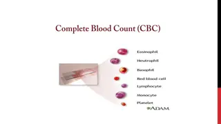
LEUKOPOIESIS the development of white blood cells (WBCs) except lymphocytes occurse in the same location as erythropoiesis Leukocytes it exist to defend the organism against nonself agents through intricate cooperation among cells. - divided into granulocytes and lymphocytes
GRANULOCYTES Contain visible granules and develop solely in the bone marrow Subdivided as granulocytes containg large granules (neutrophil, basophil, eosinophil) and granulocytes containing minute granules (monocytes)
GRANULOCYTES Can be found in four locations also called granulocyte pools: In the bone marrow b) Circulating in peripheral blood Marginating against the endothelium of blood vessels d) In the tissues a) c)
BONE MARROW POOL Has 3 functions: Proliferation b) Maturation Storage Proliferating component contain cell capable of mitotic divisions (myeloblasts, promyelocytes, and myelocytes) a) c)
Maturation component contain cells that are incapable of mitosis but not yet fully functional (metamyelocytes and bands) Storage component contains bands and polymorphonuclear leukocytes Circulating pools contain approximately 50% of total peripheral blood granulocytes levels and the other 50% in the marginating pools
STEM CELL TO MYELOBLAST Hematopoietic stem cell mature into a stem cell specific for bone marrow-derived or myeloid cells (CFU-GEMM) CFU-GEMM matures into another progenitor cell called the colony-forming unit granulocyte-monocyte/macrophage (CFU- GM) - this is controlled by by a complexof interaction humoral factors, such as interluekins and CSFs
Usually 15-20 micrometers Nucleus is delicate with prominent nucleoli Cytoplasm is meager that contains RER, developing Golgi apparatus, and the initial presence of primary or azurophilic granules The granules colors positive for enzyme MYELOPEROXIDASE Incapable of motility, adhesion, and phagocytosis < 1% in the normal bone marrow compartment MYELOBLAST
PROMYELOCYTE (PROGANULOCY TE) Size may exceed 20 micrometers Nuclear chromatin pattern may show slight clumping Nucleoli begin to fade Dominant characteristic of PRIMARY GRANULES Motility may be present 1 to 5% in the bone marrow reduced nicotinamide adenine dinucleotide phosphate oxidase and flavocytochrome B activates neutral proteinases cathepsin G, elastase, proteinases for killing to take place
<10% of the total marrow cell population Nucleus may be round to oval with flattened side near Golgi apparatus Nuclear chromatin shows clumping and nucleoli no longer visible characteristic is the production of SECONDARY OR NEUTROPHILIC granules Last stage capable of mitosis Second of the 3 major type of granules is synthesized causing dawn of neutrophilia or faint blush of pink near the Golgi apparatus NEUTROPHILIC MYELOCYTE
NEUTROPHILIC METAMYELOCYT E Result after the cessation of all active DNA synthesis Nucleus is indented Cytoplasm has collection of primary and secondary granules ( major feature of cytoplasm) The granules constitute the major component necessary to kill and degrade non self agents Incapable of reacting to chemotactic factors 13 22% of normal bone marrow differential At the end of this stage GELATINASE GRANULE is made
NEUTROPHILIC BAND (NONSEGMENTED FORM) Band shape, a transitional form because it is considered to be part of maturation and storage pools in the bone marrow and peripheral blood constitutes 40% of white blood cells but < 6% in the peripheral blood Non segmented Nucleus have uniform or parallel width Nuclear indention is less than half the width of the nucleus (horse shoe shaped) Possess full motility, adhesion properties and phagocytic ability Membrane is characterized by changes in the cytoskeleton, surface charge and presence of receptors
POLYMORPHONUCLE AR NEUTROPHIL (SEGMENTED NEUTROPHIL) Nucleus continues its indention until thin it become a lobed nucleus Nucleus is easily deformable because of active motility of the cell Polymorphonuclear means many shaped nucleus Part of storage pool in bone marrow and circulating and marginating pools 50 70% of total WBC differential Nuclei have visible segments 10 12 microns
Studies have indicated that there is a heterogeneous population of mature neutrohils Neutrophils with increased mobility, numbers of CD 15 receptors in lymphocye homing, CD21 that binds to C3 Positive for CD62 an adhesion molecule active on neutrophils and epithelial cells Performs phagocytosis (involves larger material) and pinocytosis (involves small material)
PHAGOCYTOSIS chemotactic factors cause the polymorphonuclear cell to migrate to source Neutrophils periodically determine whether the vessel endothelium is expressing surface molecules, which enhance a more firm contact (adhesion) Egress of neutrophil outside the blood circulation (diapedesis) in response to chemical gradient
It adhere to particles that initiated the attraction then pseudopods extend the around the particle, engulfing it and forming a phagosome Cytplasmic granules combine with the phagosome forming phagolysosome and dump their contents in it Primary granules contribute to proteolysis Secondary granules increase complement fixation and chemotactic response - causes degradation and detoxification of material
EOSINOPHIL MATURATION Prominent secondary granules are stained heavily with the eosin dye with Romanowsky based stains Requires IL-3, IL-5 and GM-CSF and inhibited by interferon Developed from CFU-GEMM to CFU-Eo Similar to neutrophil in the myeloblast and promyelocyte stage
In the myelocyte stage, it is distinguish from neutrophil by presence of numerous large round granules containing crystalloid compound compromising major basic protein Its granules contain proteolytic enzymes but no secretory vesicles Spends less than 1 week in the peripheral blood
Nucleus with 2 or 3 lobes connected by thin strand Large, uniform sized granules stain orange red with acidic dyes that do not obscure the nucleus 10 12 microns 1 -3 % of circulating WBCs EOSINOPHIL Charcot-Leyden crystals water soluble, needle shaped crystals as the result of eosinophil disintegration
BASOPHIL MATURATION Process of maturation from the stem cell in not well known May parallel the development of eosinophils Can be differentiated into myelocytes, metamyelocytes, bands and polymorphonuclear cells on the basis of nuclear development, although nuclei with more than two lobes are extremely rare
Large dark purple variable sized granules stain with basic dyes that obscure the nucleus Irregular shaped bilobed nucleus 8 10 microns 0 1% of circulating WBCs Have specific high affinity for the Fc region of IgE Involved in allergic inflammation and initiate localized and system anaphylaxis BASOPHILS - granules contain heparin, chondroitin sulfate, histamin, serotonine, and other vasoactive and immunomodulatory mediators
QUESTIONS 1. last stage of granulopoesis that is capable of mitosis a) Myeloblast b) promyelocyte c) myelocyte d) metamyelocyte 2. neutrophil maturation stage where it is part of both storage and maturation pool in the bone marrow a) Metamyelocyte b) band c) myelocyte d) segmented neutrophil
3. responsible for the staining of the granules of eosinophils a) Major basic protein b) peroxidase c) proteolytic enzymes d) myeloperoxidase 4. the common myeloid progenitor a) CFU-GM b) GM-CSF c) CFU-GEMM d) G-CSF 5. involved in allergic inflammation and initiate localized and system anaphylaxis a) BAND neutrophil b) basophil c) PMN d) eosinophil