Hydronephrosis and Vesicoureteral Reflux - A Comprehensive Guide
Hydronephrosis and vesicoureteral reflux are common conditions involving the urinary system. This guide covers differential diagnosis, management of physiologic hydronephrosis, distinguishing it from obstruction, and the importance of treating asymptomatic bacteriuria in pregnant women to prevent complications like acute pyelonephritis. Expert insights from Dr. Farhanul Huda, Associate Professor at AIIMS Rishikesh, provide valuable information for understanding and managing these conditions effectively.

Download Presentation

Please find below an Image/Link to download the presentation.
The content on the website is provided AS IS for your information and personal use only. It may not be sold, licensed, or shared on other websites without obtaining consent from the author. Download presentation by click this link. If you encounter any issues during the download, it is possible that the publisher has removed the file from their server.
E N D
Presentation Transcript
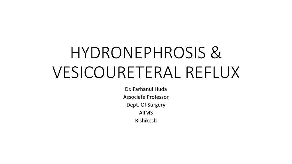
HYDRONEPHROSIS & VESICOURETERAL REFLUX Dr. Farhanul Huda Associate Professor Dept. Of Surgery AIIMS Rishikesh
A 32yearold pregnant woman presents to the ER with right sided flank pain. Renal US shows right hydronephrosis. What is the differential diagnosis?
What is the differential diagnosis? Physiologic hydronephrosis. Dilation of the upper urinary tracts occurs by the 7th week of gestation and may persist for 6 weeks postpartum. Results from both hormonal and mechanical factors. Ureteral dilation is more pronounced on the right side because of dextrorotation of the uterus, whereas the left ureter is more protected from compression by the gas filled sigmoid colon.
How do you distinguish physiologic hydronephrosis from intrinsic obstruction due to distal ureteral stone disease? Physiological hydronephrosis typically is more on the right side and generally terminates at the pelvic brim. If the hydroureteronephrosis extends below the pelvic brim, distal ureteral obstruction should be considered.
Imaging suggests physiologic hydronephrosis. How is this managed?
How is this managed? Analgesia Positioning the patient on her left side. Ureteral stent or nephrostomy tube
The flank pain resolves with conservative measures and you see the patient in clinic 2 weeks later. Her urinalysis (UA) shows bacteriuria, but she is asymptomatic. Should this be treated?
Should this be treated? Because of the risk of acute pyelonephritis. The rate of progression of asymptomatic bacteriuria to symptomatic infection is 3 4 times higher during pregnancy.
Introduction Common clinical condition. Defined as distention of the renal calyces and pelvis with urine as a result of obstruction of the outflow of urine. Can be physiologic or pathologic, acute or chronic, unilateral or bilateral. Obstructive uropathy ? Functional or anatomic obstruction of urinary flow at any level of the urinary tract. Obstructive nephropathy ? When the obstruction causes functional or anatomic renal damage.
Pathophysiology Interruption in the flow of urine Increased ureteral pressure Decreased GFR
This decline of GFR can persist for weeks after relief of obstruction. Decreased function of the nephrons. The extent of functional insult is directly related to the duration and extent of the obstruction. Brief obstruction- reversible functional changes. Chronic obstruction - profound tubular atrophy and permanent nephron loss.
Causes of U/L obstruction Extramural obstruction Tumour from adjacent structures, e.g. carcinoma of the cervix, prostate, rectum, colon or caecum Idiopathic retroperitoneal fibrosis Retrocaval ureter
Intramural obstruction Congenital stenosis, PUJO Ureterocele and congenital small ureteric orifice Stricture Neoplasm of the ureter or bladder cancer involving the ureteric orifice
Intraluminal obstruction Calculus in the pelvis or ureter Sloughed papilla in papillary necrosis due to?------
Causes of B/L Hydronephrosis congenital: posterior urethral valves; urethral atresia; acquired: benign prostatic enlargement or carcinoma of the prostate; postoperative bladder neck scarring; urethral stricture; phimosis.
Clinical features Mild pain or dull aching in the loin. Palpable kidney New onset HTN Recurrent UTIs Attacks of acute renal colic may occur with no palpable swelling. Intermittent hydronephrosis (Dietl s crisis). A swelling in the loin is associated with acute renal pain. Some hours later the pain is relieved and the swelling disappears when a large volume of urine is passed.
Investigations urine analysis Assesment of renal function USG Colour Doppler USG CT urography MR urography IVP Retrograde pyelography Isotope renography Very occasionally, a Whitaker test is indicated. A percutaneous puncture of the kidney is made through the loin and fluid is infused at a constant rate with monitoring of intrapelvic pressure.
Treatment 1. Pain management. 2. Renal drainage stenting/nephrostomy why? 3. Treat the cause 4. Anderson-Hynes pyeloplasty. 5. Endoscopic pyelolysis
INTRODUCTION Characterized by the retrograde flow of urine from the bladder to the kidneys. VUR may be associated with (UTI), HDN and abnormal kidney development (renal dysplasia). Increased risk for pyelonephritis, hypertension, and progressive renal failure. Early diagnosis and vigilant monitoring of VUR are the cornerstones of management.
CAUSES Primary causes Short or absent intravesical ureter Absence of adequate detrusor backing Lateral displacement of the ureteral orifice Paraureteral (Hutch) diverticulum Secondary causes Cystitis or UTI Bladder outlet obstruction Neurogenic bladder Detrusor instability
Pathophysiology Ureter inserts into the trigone. The intramural portion of the ureter courses into the bladder wall at an oblique angle. This intramural tunnel length to ureteral diameter ratio is 5:1. As the bladder fills with urine and the bladder wall distends and thins, the intramural portion of the ureter also stretches, thins out, and becomes compressed against the detrusor backing. This functions as a flap-valve mechanism and prevents urinary reflux.
Pathophysiology A short intramural tunnel results in a malfunctioning flap-valve mechanism and VUR. Reflux of infected urine is responsible for the renal damage. bacterial endotoxins leads to release of oxygen free radicals. These oxygen free radicals result in fibrosis and scarring of the affected renal parenchyma.
Clinical features No specific signs or symptoms unless complicated by UTI. LUTS. Palpable kidney. New onset HTN. Renal failure.
Lab studies Urinalysis and urine culture. RFT. Serum electrolytes
Imaging studies VCUG/radionuclear cystouretherography USG Nuclear scan Urodynamic studies Cystoscopy
Medical treatment Surgical treatment Surveillance
Medical management Administering long-term suppressive antibiotics Correcting the underlying voiding dysfunction (if present) Conducting follow-up radiographic studies (eg, VCUG, nuclear cystography, DMSA scan) at regular intervals
Surgical treatment Grade III or more reflux. Failure of medical management. Ureteral reimplantation Endoscopic treatment: The principle of the procedure is to inject, under cystoscopic guidance, a biocompatible bulking agent underneath the intravesical portion of the ureter in a submucosal location. The bulking agent elevates the ureteral orifice and distal ureter in such a way that the lumen is narrowed, preventing regurgitation of urine up the ureter but still allowing its antegrade flow.
A 50 year old female is admitted with abdominal pain and anuria. Radiological studies reveal bilateral impacted ureteric stones with hydronephrosis. Urine analysis showed RBCs with pus cells in urine. Serum creatinine level was 16 mg / dl and blood urea level was 200 mmol/L; which of the following should be the immediate treatment. a. Lithotripsy b. Ureteroscopic removal of stones c. J stent drainage d. Hemodialysis