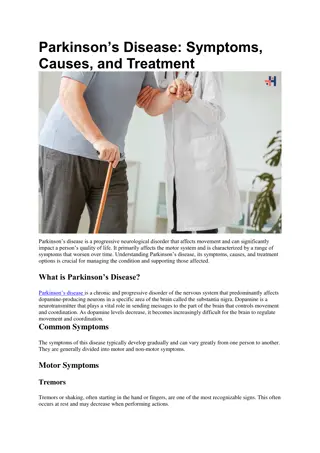
Hoarseness: Causes, Symptoms, and Treatment
Hoarseness is roughness of voice caused by various factors such as inflammation, structural issues, and muscle tension imbalance. Learn about the clinical approach, common causes like acute laryngitis, and supportive measures for treatment.

Download Presentation

Please find below an Image/Link to download the presentation.
The content on the website is provided AS IS for your information and personal use only. It may not be sold, licensed, or shared on other websites without obtaining consent from the author.If you encounter any issues during the download, it is possible that the publisher has removed the file from their server.
You are allowed to download the files provided on this website for personal or commercial use, subject to the condition that they are used lawfully. All files are the property of their respective owners.
The content on the website is provided AS IS for your information and personal use only. It may not be sold, licensed, or shared on other websites without obtaining consent from the author.
E N D
Presentation Transcript
Anatomy of the Spinal Cord Important Doctors Notes Notes/Extra explanation Lecture (2) Editing File Please check our Editing File 436 { { } }
Objectives At the end of the lecture, the students should be able to: Describe the external anatomy of the spinal cord. Describe the internal anatomy of the spinal cord. Describe the spinal nerves: formation, branches and distribution via plexuses. Define Dermatome and describe its significance. Describe the meninges of the spinal cord. Define a reflex and reflex arc, and describe the components of the reflex arc. The first 4 slides of the boys lecture were not included since they are a review of the first lecture.
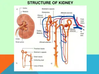
Spinal Cord Spinal Cord o The main pathway for information connecting the brain and peripheral nervous system. o An elongated, almost cylindrical structure, about the thickness of the little finger. o It is suspended in the vertebral canal & surrounded by the meninges and cerebrospinal fluid (CSF). o In adults, its Length is approximately 45 cm. o The primary function of spinal cord is a transmission of neural signals between the brain and the rest of the body. Sensory Motor Local reflexes
Spinal Cord Spinal Cord o Extends from foramen magnum to L1-L2 (intervertebral disc). (boys slides: until second lumbar vertebra L2) o In children it extends to L3 because their vertebral column is smaller/shorter. o Continuous above with the medulla oblongata. o The tapered inferior end forms conus medullaris*, which is connected to the coccyx by a non-neuronal cord called filum terminale (its not considered a part of the spinal cord.). o Gives rise to 31 pairs of spinal nerves. Extra ( *cone like ( (its enlarged because it supplies the lower limbs and it is the place anestheics are injected during child birth). Extra
Image result for youtube Spinal Cord Spinal Cord 07:22 o The spinal cord is a Segmented structure, has 8 Cervical 12 Thoracic 5 Lumbar 5 Sacral 1 Coccygeal segments o Not uniform in diameter, (not the same diameter throughout) o Has two enlargements: Cervical enlargement: supplies upper limbs Lumbosacral enlargement: supplies lower limbs o The bundle of spinal nerves extending inferiorly from lumbosacral enlargement and conus medullaris surround the filum terminale and form cauda equina (because of its resemblance to a horse's tail) The enlargements to supply the needs of upper and lower limbs *Recall: End of spinal cord: conus medullaris. End of spinal nerves: cauda equina.
You can know the orientation by: 1- dorsal root ganglion 2- anterior median fissure Fissure > sulcus Spinal Cord Cross Section Cross Section Ventral = Anterior Dorsal = Posterior o The spinal cord is incompletely divided into two equal parts, anteriorly by a short, shallow median fissure (thicker) and posteriorly by a deep narrow septum, the posterior median septum (sulcus). o Composed of grey matter in the centre surrounded by white matter supported by neuroglia.* o The arrangement of grey matter resembles the shape of the letter H, having two posterior, two anterior and two lateral horns/columns. (not all grey matter has lateral horns) * * * o Commissures: connections between left and right halves Gray with central canal in the center White (both will be discussed later) o Roots: spinal nerves arise as rootlets then combine to form roots: Dorsal (posterior) root has a ganglion and Ventral (anterior). Two roots merge laterally and form the spinal nerve. *Don t be confused: The brain Spinal cord Cortex outer layer Gray matter White matter Medulla inner layer White matter Gray matter
Grey Matter Grey Matter Image result for youtube 07:46 o Consists of (1) nerve cell bodies and their processes, (2) neuroglia, and (3) blood vessels o The nerve cells are multipolar and are of three main categories: Ventral = Anterior Dorsal = Posterior 1. Sensory neurons (Tract cells), which receive impulses from the periphery of the body and whose axons constitute the ascending fasciculi of the white matter, are located in the Dorsal horns. 2. Lower motor neurons, which transmit impulses to the skeletal muscles, are located in the ventral horns (similar neurons in the lateral horn are the preganglionic neurons of the autonomic system) 3. Interneurons (connector neurons): linking sensory and motor neurons, at the same or different levels, which form spinal reflex arcs. (only in spinal cord not brain)
Spinal Grey Matter Neuronal Architecture Neuronal Architecture Dorsal horn: 1 7 Ventral horn: 8, 9 Central horn: 10 oCells of the same type are clustered into groups, which occur in long columns oIn transverse section, these columns appear as layers, especially within the dorsal horn oThese layers are called the Laminae of Rexed, that are numbered consecutively by Roman numerals, starting from the tip of the dorsal horn and moving ventrally into the ventral horn. oThe rexed laminae comprise a system of ten layers of grey matter (I-X), identified in the early 1950s by a Swedish neuroscientist. Extra Extr a
Dorsal Horn Nerve Cell Groups Nerve Cell Groups 5 main groups 1. Substantia gelatinosa 2. Nucleus proprius 3. Nucleus dorsalis (Clark s column, nucleus thoracis) 4. Visceral afferent nucleus 5. Marginal zone (posterior marginals)* 1 2 3 4 * Only in boys slides
Dorsal Horn Nerve Cell Groups Nerve Cell Groups The doctor said: you should know the correspondence of each lamina. For example, lamina 2 corresponds to substatia gelationsa Marginal zone (posterior marginals) Rexed Laminae Location Composed of Extends Afferents I (1) At the tip of dorsal horn Important for relaying pain and temperature sensation to the brain. - - Substantia Gelatinosa Rexed Laminae Location Composed of Extends Afferents II (2) apex of the posterior/ dorsal horn large neurons throughout the length of spinal cord dorsal root fibers concerned with pain, temperatureand crude touch Nucleus Proprius Rexed Laminae Location Composed of Extends Afferents dorsal root fibers concerned with fine touch (senses of position & movement (proprioception) and two point discrimiation & vibration) * IV (4) anterior to substantia gelatinosa/ located in the neck of dorsal horn large neurons throughout the length of spinal cord *There is variation in this point between boys and girls slides, This is the correct information according to Snell s Clinical Neuroanatomy
Dorsal Horn Nerve Cell Groups Nerve Cell Groups Nucleus Dorsalis (Clark s column, Nucleus thoracis) Rexed Laminae Location Composed of Extends Afferents base of dorsal horn/ the most dorso-medial nuclei from C8 to L3-4 segments VII (7) mostly large neurons dorsal root fibers concerned with information from muscle spindles and tendon organs. *Associated with proprioceptive endings *Relays unconscious proprioceptive information to brain Visceral Afferent Nucleus Rexed Laminae Location lateral to nucleus dorsalis Composed of mostly of medium size neurons Extends Afferents Visceral afferents VII (7) from T1 to L3 segments
Only in boys slides *The doctor noted that we only required to know the location of each nucleus in previous slide in which lamina Rexed laminae Lamina I: Lamina I: o tip of the dorsal horn o cells respond to noxious or thermal stimuli o sends information to the brain by the contralateral spinothalamic tract o corresponds to the marginal zone Lamina II: Lamina II: o Involved in sensation of noxious and non-noxious stimuli, and modulating sensory input to contribute to the brain s interpretation of incoming signals as painful, or not. o Sends information to Lamina III and IV o Corresponds to substantia gelatinosa. Lamina III: Lamina III: o Involved in proprioception and sensation of light touch. o Cells in this layer connects with cells in layers IV, V and VI. o Partially corresponds to nucleus proprius Lamina IV: Lamina IV: o Involved in non-noxious sensory information relay and processing. o Cells connect with those in lamina II o Partially corresponds to nucleus proprius Lamina V: Lamina V: o Relays sensory, including nociceptive (potentially painful), information to the brain via the contralateral and spinothalamic tracts o Receives descending information from the brain via the corticospinal and rubrospinal tracts. Lamina VI: Lamina VI: o Contains many small interneurons involved in spinal reflexes o Receives sensory information from muscle spindles (involved in proprioception). o Sends information to the brain via ipsilateral spinocerebellar pathways Lamina VII: Lamina VII: o Large, heterogenous zone that varies through the length of the spinal cord. o Receives information from Lamina II to VI, and from viscera o Relays motor information to the viscera o Gives rise to cells involved in the autonomic system. o Dorsal nucleus of Clarke is part of Lamina VII Lamina VIII: Lamina VIII: o Varies depending on spinal cord level, but is most prominent in cervical and lumbar enlargements. o Cells are involved in modulating motor output to skeletal muscle. Lamina IX: Lamina IX: o Size and shape varies between spinal cord levels. o Distinct groups of motor neurons that innervate skeletal muscle. Lamina X: Lamina X: o Surrounds the central canal the grey commissure. o Axons decussate (cross over) from one side of the spinal cord to the other.
Ventral Horn Nerve Cell Groups Nerve Cell Groups The ventral horns contain: 1. Motor neurons, also called lower motor neurons (the upper motor neurons are in the brain). 2. Interneurons, the (Renshaw cells), whose branched axons form inhibitory synaptic junctions on motor neurons
Ventral Horn 1 1. Motor Neurons . Motor Neurons There are two types of motor neurons in ventral horn: o Large multipolar cells Numerous Axons pass out in the ventral roots of spinal nerves as alpha efferents Innervate extrafusal muscle fibers o Smaller multipolar cells Less numerous Axons pass out in the ventral roots of spinal nerves as gamma efferents Innervate intrafusal muscle fibers of neuromuscular spindles Both alpha and gamma motor neurons are under the influence of descending pathways (upper motor neurons) from brain.
Ventral Horn 1. Motor Neurons 1. Motor Neurons In the the ventral horn motor neurons are organized in 3 groups: Medial Central Lateral present in most segments present in some segments: cervical (phrenic C3-5, spinal accessory C1-6) and lumbosacral (L2-S1) present in cervical and lumbosacral segments Innervate muscles of Neck and Trunk (including intercostal and abdominal muscles) smallest innervates muscles of the Limbs Neurons supplying flexor muscles are located dorsal to neurons for extensor muscles.
Lateral Horn Nerve Cell Groups Nerve Cell Groups Small column composed of small neurons extend from: T1 to L2-3 segments, give rise to pre-ganglionic sympathetic fibers (thoracolumbar). S2-4 segments, give rise to preganglionic parasympathetic fibers (craniosacral). Interomediolateral Nucleus(IMN): Located in the intermediate column and lateral horn. Relays sensory information from viscera to the brain , and autonomic signals from the brain to the visceral organs.
Image result for youtube White Matter White Matter 09:14 o Consists of mixture of nerve fibers, neuroglia and blood vessels. o White color is due to high proportion of myelinated nerve fibers. o Arranged in columns/funiculi; anterior, posterior and lateral. o The nerve fibers are arranged as bundles, running vertically through the cord. A group of nerve fibers (axons) that share a common origin, termination and function form a tract or fasciculus. o These tracts are formed by (1) sensory nerve fibers ascending to the brain, (2) motor nerve fibers descending from the brain and (3) fibers of connector neurons. o Depending on their function, the spinal tracts are divided into ascending and descending tracts. o Tracts are often named according to their points of origin and destination, e.g. spinothalamic, corticospinal. Funiculi:a bodily structure suggesting a cord; especially : a bundle of nerve fibers Fasciculus: a small or slender bundle (as of nerve fibers)
Central Canal o The central canal is a cerebrospinal- filled space that runs longitudinally through the entire length of the spinal cord. o Lined by ependyma (ciliated columnar epithelium) (important) o Continuous with the ventricular system of the brain o Superiorly opens into the 4thventricle o Inferiorly in the conus medullaris, it expands into the fusiform terminal ventricle and terminates below at the root of filum terminale. Extra (important) Extra Extra
Spinal Cord Commissures Commissures Grey commissure: o A transverse bridge of grey matter connecting the anterior and posterior gray horns on each side o Is pierced by the central canal that divides it into anterior and posterior parts White commissure Grey commissure White Commissure: o Lies ventral (anterior) to the gray commissure o Mainly contains decussating* nerve fibers *decussating: ( ) cross or intersect each other to form an X Extra
Thoracic Cervical Spinal Cord Spinal Cord Regional Differences o Although the general pattern of gray matter is the same throughout spinal cord, regional differences are apparent in transverse sections Sacral Lumbar o The amount of white matter increases in a caudal-to-cranial direction because fibers are added to ascending tracts o The gray matter is increased in volume in cervical & lumbosacral enlargements for innervation of upper & lower limbs o The lateral horn is characteristics of thoracic and upper lumbar segments Extra
Spinal Meninges Spinal Meninges Image result for youtube 07:07 o Three connective tissue membranes* surround spinal cord and brain: Dura mater: tough outer layer, continuous with epineurium of the spinal nerves Arachnoid mater: thin and wispy membrane deeper to dura mater Pia mater: delicate membrane bound tightly to surface of brain and spinal cord and carries blood vessels. Forms the filum terminale, which anchors spinal cord to coccyx and the denticulate ligaments that attach the spinal cord to the dura mater o Spaces: Epidural: Contains blood vessels, areolar connective tissue and fat. (between dura and bone: skull/vertebra) Subdural: a potential cavity between the dura and arachnoid mater, contains a small volume of serous fluid. Subarachnoid: Contains cerebrospinal fluid (CSF) and blood vessels within web-like strands of arachnoid tissue. (between arachnoid and pia mater) CSF can be collected from this space for diagnostic purposes. P A D Dura Outside Pia Inside
Spinal Nerves Spinal Nerves o Thirty-one pairs of spinal nerves. o First pair exit vertebral column between skull and atlas, last four pair exit via the sacral foramina and others exit through intervertebral foramina o Eight pair cervical, twelve pair thoracic, five pair lumbar, five pair sacral, one pair coccygeal o Each spinal nerve arises as rootlets which then combine to form dorsal (posterior) purely sensory & ventral (anterior) purely motor Roots. o Two roots merge laterally and form the spinal nerve. o Dorsal (posterior) root has a ganglion (dorsal root/sensory ganglion) that contains the cell bodies of the sensory neurons. o Each spinal nerve then divides into a MIXED smaller dorsal and a larger ventral Ramus Rootlet Root Spinal Nerve Rami Dorsal Dorsal Purely Sensory Ventral Ventral Purely Motor Mixed Extra
Spinal Nerves Branches Branches Ventral = Anterior Dorsal = Posterior o Dorsal Rami innervate: Deep muscles of the trunk responsible for movements of the vertebral column Skin near the midline of the back. o Ventral Rami: In the thoracic region form intercostal nerves that innervate the intercostal muscles and the skin over the thorax Remaining ventral rami form five plexuses *: C1 - C4= Cervical plexus C5 - T1= Brachial plexus L1 - L4= Lumbar plexus L4 - S4= Sacral plexus S5 & Co= Coccygeal plexus** (intermingling of nerves) *see the next slide **There is variation in this point between boys and girls slides, This is the correct information according to Grey s Anatomy for Students
Spinal Nerves Branches Branches The spinal nerves are connected to sympathetic chain of ganglia by communicating rami. Extra
Dermatomes Dermatomes o Dermatome is a segment of skin supplied by a specific segment of the spinal cord (segmental spinal nerve), i.e, one nerve. o Cutaneous areas supplied by adjacent spinal nerves overlap. There is therefore little or no sensory loss after interruption of a single spinal nerve or dorsal root Reflex & Reflex Arc Reflex & Reflex Arc o A reflex is a rapid, involuntary, predictable, stereotyped pattern of response brought by a sensory stimulus. o The neural pathway mediating the reflex actions is called reflex arc.
Components of a Reflex Arc Action potentials produced in Interneuron Sensory receptor Sensory neuron Effector organ which responds with a reflex Motor neuron Variety of Reflexes o Some integrated within spinal cord; some within brain o Some involve excitatory neurons yielding a response; some involve inhibitory neurons that prevent an action o Higher brain centers can influence, suppress, or exaggerate reflex responses Only on the girls slides
Only in boys slides CLINICAL SIGNIFICANCES Spinal cord injuries can be caused by trauma to the spinal column (stretching, bruising, applying pressure, severing). Usually, victims of spinal cord injuries will suffer loss of feeling in certain parts of their body. In milder cases, a victim might only suffer loss of hand or foot function. More severe injuries may result in paraplegia, tetraplegia (also known as quadriplegia), or full body paralysis below the site of injury to the spinal cord. Spinal shock and neurogenic shock can occur from a spinal injury. Spinal shock is usually temporary, lasting only for 24 48 hours, and is a temporary absence of sensory and motor functions. Neurogenic shock lasts for weeks and can lead to a loss of muscle tone due to disuse of the muscles below the injured site.
MCQs MCQs 1. The tapered inferior end of the spinal cord forms: A- filum terminale B- conus medullaris C- cauda equina D- denticulate ligaments Answer: B 5. Dorsal root fibers concerned with information from muscle spindles are found in which laminae? A- II B- IV C- VI D- VII Answer: D 2. Filum terminale is formed from: A- dura mater B- arachnoid mater C- pia mater D- spinal nerves Answer: C 6. In which segment of the spinal chord can we find lateral horn: A- cervical B- thoracic C- coccygeal Answer: B 3. If a cross section of the spinal cord is taken from C8 which of the following nerve cell group will be missing? A- Substantia gelatinosa B- Nucleus proprius C- Nucleus dorsalis D- Visceral afferent nucleus Answer: D 7. A plexus is made of: A- dorsal root B- dorsal rami C- ventral root D- ventral rami Answer: D 8. Which of the following is continuous with epineurium of the spinal nerves? A- dura mater B- arachnoid mater C- pia mater Answer: A 4. Renshaw cells are present in which horn? A- Lateral B- Ventral C- Dorsal Answer: B
MCQs MCQs 9. : the Brachial plexus is from to : A- C1-C4 B- C5-T1 C- L5-Co D- S4-L$ Answer: B 10. The spinal cord has: A- 30 pairs of spinal nerves B- 30 spinal nerves C- 31 pairs of spinal nerves D- 31 spinal nerves Answer: C SAQ SAQ Q1: Mention the location of Nucleus Dorsalis in the spinal cord ? Answer: from C8 to L3-4 segments in the base of the dorsal horn. Q2: Define Reflex Arc . Answer: It s is a rapid, involuntary, predictable, stereotyped pattern of response brought by a sensory stimulus.
Good luck Special thank for team436 Team Leaders: Faisal Fahad Alsaif Rawan Mohammad Alharbi Team Members: Abdulaziz Aldukhayel Abdulrahman Alduhayyim Rinad Alghoraiby Rawan Mishal References: 1.Girls & Boys Slides 2.Greys Anatomy for Students 3.TeachMeAnatomy.com Twitter.com/Anatomy437 Anatomyteam.437@gmail.com