Hematocrit (PCV) Test: Importance and Procedure

Hematocrit (PCV) is crucial for assessing anemia or polycythemia. It measures the percentage of blood volume occupied by red blood cells. This test is accurate and simple, providing valuable information for diagnosing various conditions. Normal values, causes of increased or decreased PCV, and the procedure for conducting the test are discussed.

Download Presentation

Please find below an Image/Link to download the presentation.
The content on the website is provided AS IS for your information and personal use only. It may not be sold, licensed, or shared on other websites without obtaining consent from the author.If you encounter any issues during the download, it is possible that the publisher has removed the file from their server.
You are allowed to download the files provided on this website for personal or commercial use, subject to the condition that they are used lawfully. All files are the property of their respective owners.
The content on the website is provided AS IS for your information and personal use only. It may not be sold, licensed, or shared on other websites without obtaining consent from the author.
E N D
Presentation Transcript
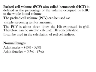
Hematocrit Hematocrit: is the proportion of blood volume that is occupied by erythrocytes (the ratio of red blood cells to the whole blood volume). It is usually expressed as a percentage.
Relevance Measurement of hematocrit (Hct) or packed cell volume (PCV) is the most accurate and simplest of all tests for detecting the presence and degree of anemia or polycythemia. In comparison, hemoglobin estimation is less accurate, and RBC count far less accurate.
Relevance Also, if Hb, RBC count, and PCV are determined at the same time, various absolute corpuscular values (e.g., volume and Hb content of a single red cell) of a person can be determined. These values help in the laboratory diagnosis of the type of anemia in a person.
Normal values The average levels and their ranges are as follows: Males: (38-50) %, (44 %). Females: (36 45) %, (42 %).
Increased PCV 1. Polycythemia vera (primary polycythemia) 2. Secondary polycythemia 3. Polycythemia due to hemoconcentration 4. Physiological polycythemia
Decreased PCV 1. Medications 2. Genetic conditions 3. Bleeding 4. Medical conditions 5. Nutritional deficiencies 6. Physiological conditions
Apparatus and materials 1. Heparinized capillary tubes 2. Microhematocrit centrifuge 3. Microhematocrit reader 4. Modeling clay sealant 5. Blood lancet, sterile cotton/ gauze swabs, 70% alcohol
Procedure 1. Puncture the skin of the finger for collecting blood, directly fill 2/3 of heparinized microhaematocrit tube with blood. 2. Seal one end of the tube with clay.
Procedure 3. Place the filled tube in the microhematocrit centrifuge, with the plugged end away from the center of the centrifuge and centrifuge at a preset speed of 10,000 to12,000 rpm for 5 minutes.
Procedure 4. Place the tube in the microhematocrit reader. 5. Read the hematocrit by following the manufacturer s instruction on the microhematocrit reading device.
Observations and results Note that the blood has been separated into 3 layers: 1. Atall upper layer of clear plasma Amber or pale yellow-colored. It should not be pink or red which would indicate hemolysis of red cells in the sample. If there is hemolysis, the test must be repeated on a fresh sample.
Observations and results 2. Agreyish layer White, thin layer (about 1 mm thick), so called buffy layer . Consisting of platelets above and leukocytes below it. 3. A tall bottom layer of red cells which have been closely packed together.
Observations and results Read the hematocrit as follows; 1. Place the bottom of the tube along the red line below the scale. 2. Be sure that the place where the white hematocrit sealer meets the RBC s is on the line at the bottom of the scale itself.
Observations and results 3. Move the tube along those lines until the top of the plasma intersects the top line of the scale 4. The value of the hematocrit is read on the line where the RBC s meet the plasma.
PRECAUTIONS 1. Use the recommended amount of EDTA as the anticoagulant. There should be no clotting or hemolysis of the blood. 2. The test should be done within 6 8 hours of collection of the sample.