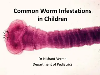
Giant Intestinal Roundworms: Ascaris lumbricoides
Ascaris lumbricoides, also known as the Giant Intestinal Roundworm, is a common parasitic nematode infecting the human intestines, especially prevalent in underdeveloped regions with poor sanitation. Found in the small intestines, this worm can cause significant health issues. Learn about its morphology, geographical distribution, life cycle, and impact on human health.

Download Presentation

Please find below an Image/Link to download the presentation.
The content on the website is provided AS IS for your information and personal use only. It may not be sold, licensed, or shared on other websites without obtaining consent from the author.If you encounter any issues during the download, it is possible that the publisher has removed the file from their server.
You are allowed to download the files provided on this website for personal or commercial use, subject to the condition that they are used lawfully. All files are the property of their respective owners.
The content on the website is provided AS IS for your information and personal use only. It may not be sold, licensed, or shared on other websites without obtaining consent from the author.
E N D
Presentation Transcript
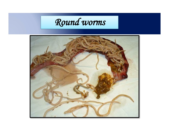
Round Round worms worms
INTRODUCTION Ascaris lumbricoides is the largest nematode (roundworm) parasitizingthe human intestine. Ascaris lumbricoides is an intestinal worm found in the small intestine of man(mainly in the jejunum and upperpart of theileum). They are more commonin children than inadults As many as 500 to 5000 adult worms may inhabit a single host.
Common name:- Giant Intestinal round worms. Disease:-Ascariaisis Host :- The human is intermediate and final host. Location in Definitive host :- the adult worm: in small intestines . larva: in lung . Infective stage :- embryonated eggs
Geographicaldistribution Worldwide High prevalence in underdeveloped countries that havepoor sanitation(partsof Asia,South America andAfrica) Occurs during rainy months, tropicaland subtropicalcountries Even occurs in rural areas in the UnitedStates
MORPHOLOGY It is a elongated, cylindricaland tapering at bothends. Sexes areseparate The female is longer than male25 40 cm long, 4-6 mmin diameter. Male is smaller being 15-30cm long, 2-4 mm indiameter. The posterior end of male is curvedventrallyintheformofa hook The digestive and respiratory organs of the worm float inside the body cavity possessing atoxic fluid known asascaron
The Mouth Parts The mouth opens at theanterior end. It is surrounded by threefinely toothed lips. The lips are one dorsal andtwo ventrolateral. These lips bear sensorystructures called labialpapillae
A mature female A. lumbricoides lays enormous number ofeggs (nearly 2,00,000 eggs daily) which are passed in thefaeces There are two kinds ofthe eggs. They are fertilized eggs, and unfertilized eggs We usually describe an egg in 5 aspects: size, color,shape, shell andcontent Decorticated eggs: Both fertilized and unfertilized eggs sometimes may lack their outer albuminous coats andare colorless
Fertilized Egg Broad oval in shape,brown in color, anaveragesize60 45 m. The shell is thicker and consists of chitinous layer, and mammillated albuminous coat stained brown by bile. The content is a fertilizedovum. There is a new-moon(crescent) shaped clear space at the each end insidetheshell.
Unfertilizedegg Narrower and longer and measure 90 m in length and 55 m in breadth They are bile stained and brown in colour The chitinous layer and albuminous coat are thinner and irregular than those of the fertilizedeggs The content is made of atrophied ovum many refractable granules of various size. Heaviest of all the helminthiceggs small suurounded by
Decorticatedeggs Both fertilized and unfertilized eggs sometimes may lack their outer albuminous coats and arecolorless.
Modes oftransmission Occurs mainly via ingestion of water or food (raw vegetablesorfruitsinparticular)contaminatedwithA. lumbricoideseggs. Occasionally inhalation of contaminateddust Children playing in contaminated soil may acquirethe parasite from theirhands Enhanced by the fact that individuals can be asymptomatically infected and continues to shedeggs for years
Stage I: Eggs in faeces Sexually mature female produces as many as 200,000 eggs per day, which are shed along with faeces in unembryonated form. They are non infective. Stage II: Development in soil Embryonation occurs in soil as optimum temperature of 20-25C with sufficient moisture and O2. Infective larva develops within egg in about 3-6 weeks. Stage III: Human infection and liberation of larvae Human get infection with ingestion of embryonated egg contaminated food and water Within embryonated state inside egg, first stage larvae develops into second stage larvae. This second stage larvae is known as Rhabtitiform larvae Second stage larvae is stimulated to hatch out by the presence of alkaline pH in small intestine and solubilization of its outer layer by bile.
Stage IV: migration of larvae through lungs Hatched out larvae penetrates the intestinal wall and carried to liver through portal circulation. It then travels via blood to heart and to lungs by pulmonary circulation within 4-7 days of infection. The larvae in lungs molds twice, enlarge and breaks into alveoli.
Stage V: Re-entry to stomach and small intestine From alveoli, the Larvae then pass up through bronchi and into trachea and then swallowed. The larvae passes down the oesophagus to the stomach and reached into small intestine once again. Small intestine is the normal habitat of Ascaris and it colonises here. Within intestine parasite molds twice and mature into adult worm. Sexual maturation occurs with 6-10 weeks and the mature female discharges its eggs in intestinal lumen and excreted along with faeces, continuing the life cycle. The life span of parasite is 12-18 months
Pathogenesis Infection of A. lumbricoides in man is known as Ascariasis. There are two phase in ascariasis. Phase I: migrating larvae The migrating larvae causes pathological lesions. The severity of lesions depends upon the sensitivity of host, nutritional status of host and number of migrating larvae. During migration and molding through lungs, larvae may causes pneumonia with low grade fever, cough and other allergic symptoms.
Phase II: Adult worm Few worm in intestine produce no major symptoms and but some time give abdominal pain especially in children. The adult worm produce trauma in host tissue and the wandering adults may block the appendical lumen or common bile duct and even small intestine. Large number of adult worms affects the nutritional status of host by robbing the nutrition leading to malnutrition and growth retardation in children. The metabolites of living or dead worm are toxic and immunogenic. lumbricoides also produces various allergic toxin, which manifests fever, conjunctivitis and irritation.
Clinical manifestation: Most of the Ascaris infection is asymptomatic. Symptomatic ascariasis; two types: Intestinal Ascariasis Pulmonary Ascariasis 1. Intestinal ascariasis; Nausea Vomiting Colicky abdominal pain Abdominal distention Weight loss and diarrhea Malbasorption of nutrition Growth retardation Heavy worm in children leads to intussusception and total obstruction Complications:Appendicitis, Biliary colic and perforation of bile duct, Hepatomegaly 2. Pulmonary ascariasis; Transient eosinophilic pneumonitis (loeffler s disease); elevated IgE Bronchospasm Dyspnea and wheezing Fever Non-productive cough and chest pain
Laboratorydiagnosis Done by followingmethods 1. Parasiticdiagnosis a) Demonstration of adultworm b) Demonstration ofeggs c) Demonstration oflarvae 2. Serodiagnosis 3. Eosinophilia and Ultrasonography and CT scan
Demonstration of adultworms Worm may be passed through anus, mouth, nose andrarely through ear Barium meal may occasionally reveal the presence ofadult worms in the smallintestine Demonstration ofeggs Eggs may be detected in stool orduodenal bile aspirate by direct microscopyor after concentrationof faeces Eggs may not be seen if only male worms arepresent
Demonstraion oflarvae Ascarislarvaemaybedetectedinthesputumduringthestage ofmigration 2.Serodiagnosis Ascaris antibody can be detected by indirecthaemagglutination (IHA) And immunofluorescence antibody (IFA)test Thesetestsareusefulforthediagnosisofextraintestinal ascariasis like Loeffler ssyndrome 3.Eosinophilia It is seen in larval invasionstage
Treatment Pyrantel pamoate, in a single dose of 11mg per kilogram body weight (maximum 1gm) Mebendazolein adoseof 100mgtwice daily for 3 days,and piperazine citrate in adoseof 75mgper kgbody weight daily for 2days
PREVENTION Keeping good sanitation conditions is the only way to prevent the infection of Ascaris. Pollution of soil with human faeces should be avoided. Vegetable should be thoroughly washed in a mild solution of Pottasium permanganate and properly cooked before use. Finger nails should be regularly cut to avoid the collection of dirt and eggs below them. Hands should be properly washed with some antiseptic soap before touching edibles or eating.
Prophylaxis Ascariasis can be prevented by Proper disposal of humanfaeces Avoidance of eating raw vegetables andsalads Periodic treatment with an effective anthelminthic, in communities thatlack sanitary facilities
LarvaMigrans This is a term used to describe human infections with helminth larvae, which arenot adapted to humanbeings The condition is usually caused by animal parasites, man being an abnormal host,these larvae are not able to reach the normal habitat and keep wandering in the abnormal host (man), hence, known as larvamigrans
Divided into 2types 1) Cutaneous larva migrans (CLM) also known as creeping eruption 2) Visceral larva migrans(VLM)
Common points between CLM andVLM Man always acquires the infection asan accidentalhost The causative agents are usually zoophilic helminths The host mounts an inflammatory response directed against somatic antigens ofparasites Both diseases affect primarily thechildren Both are widespread in tropical andtemperate countries of theworld