Fellow Radiograph Template
In the field of implant dentistry, diagnostic radiographs play a crucial role in assessing bone levels and overall treatment success. This content outlines the specific radiographs required, including presurgical, postsurgical, and completed case radiographs, as well as additional imaging for grafting cases. Follow the guidelines set by the American Academy of Implant Dentistry to ensure diagnostic quality and minimal distortion in the radiographs. Stay informed about the credentialing standards and best practices to advance the science and practice of implant dentistry.
Download Presentation

Please find below an Image/Link to download the presentation.
The content on the website is provided AS IS for your information and personal use only. It may not be sold, licensed, or shared on other websites without obtaining consent from the author.If you encounter any issues during the download, it is possible that the publisher has removed the file from their server.
You are allowed to download the files provided on this website for personal or commercial use, subject to the condition that they are used lawfully. All files are the property of their respective owners.
The content on the website is provided AS IS for your information and personal use only. It may not be sold, licensed, or shared on other websites without obtaining consent from the author.
E N D
Presentation Transcript
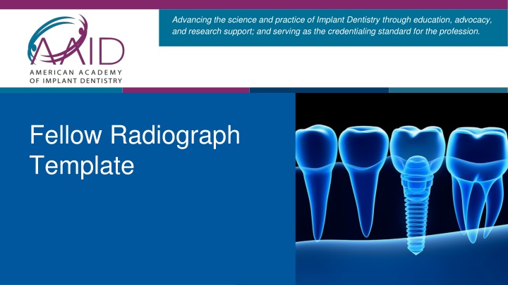
Advancing the science and practice of Implant Dentistry through education, advocacy, and research support; and serving as the credentialing standard for the profession. Fellow Radiograph Template
RADIOGRAPHS REQUIRED: All radiographs must be of diagnostic quality and have minimal distortion, and bone levels must be obvious: 1. 2. Presurgical panograph or a full-mouth radiographic series. Post-surgical (within one week of surgery) panograph or a post surgical periapical radiograph for a single-tooth-implant Post-prosthetic (with prosthesis or bar superstructure in place); either panographic or periapical radiographs are acceptable Completed case radiograph (taken within 12 months of the candidate s oral/case examination date.) 3. 4. Grafting cases only: two (2) additional radiographs are required: 1. Cross sectional cone beam radiograph of the augmentation site both BEFORE the augmentation s placement. 2. Pre-prosthetic cross sectional cone beam radiograph of augmentation site that shows 3 mm gain in bone for onlay grafts 5 mm gain in bone for sinus grafts 2023 | American Academy of Implant Dentistry
Candidate Number: {Candidate number} Patient: {ABC} Case Type: {Case Type} 2023 | American Academy of Implant Dentistry
Candidate Number: {Candidate number} Insert Radiograph View: {View} Date Radiograph taken: {01/01/2000} Patient Initials: {ABC} 2023 | American Academy of Implant Dentistry
