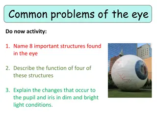
Eye Disorders in Veterinary Science

 
The eye
The eye
Miscellaneous
Miscellaneous
 
Gulshan Kumar
Gulshan Kumar
MVSc, PhD
MVSc, PhD
 
college of veterinary science & animal husbandry, duvasu, Mathura (UP)
 
Amaurosis
Amaurosis
 
is 
is 
blindness without any apparent lesion in the eye. It may be temporary or permanent. Possible
causes are toxaemia, lesions in the brain, etc. (Note: A temporary form of amaurosis is sometimes seen in
cattle due to deficiency of vitamin A which can be corrected by administration of vitamin A.)
Refraction of the eye: 
Parallel rays: The amount of divergence of light rays falling on a given area is inversely
proportionate to the distance from the source of light. When the distance is 20 feet or more, the divergence
is so slight that the rays can be considered as parallel
.
.
Emmetropia (Normal sight):
When the refraction of the eye is normal, parallel rays coming into the eye in a
condition of rest, are focused exactly on the retina. This condition is known as emmetropia.
Ametropia 
Ametropia 
is a term used to denote a condition of abnormal refraction of the eye due to hypermetropia,
myopia, or astigmatism, in which parallel rays are focused either in front or behind the retina.
Hypermetropia(Hyperopia; Long sight; Far sight
): Hypermetropia is a condition of abnormal refraction of the eye
in which parallel rays come to a focus behind the retina. This type of ametropia is caused if the axis of the
eyeball is too short or if the refractive power of the eye is too weak.
Myopia(Short sight; Near sight): 
Myopia is a condition of abnormal refraction of the eye in which parallel rays
get focused in front of the retina. This may happen either due to the axis of the eyeball being too long or
due to the refractive power of the eye being too strong. (In this condition the eye is able to see clearly only
objects very close to it.)
 
college of veterinary science & animal husbandry, duvasu, Mathura (UP)
 
Astigmatism: 
Astigmatism: 
When the refraction through several meridians of the eye is different, the
condition is called astigmatism. Agtigmatism may be caused by irregularities in the cornea or
the lens. Astigmatism causes blurred vision. (Note: A certain degree of astigmatism is normally
present in the horse.)
 
Disease of the vitreous, retina, choroid and optic nerve/posterior segment
The image obtained in the ophthalmoscope while viewing the posterior segment is called fundus
and it comprises of optic disc, retinal vasculature, and a semitransparent neurosensory retina.
Through this, structures like retinal pigment epithelium chorioid or tapetum and posterior sclera
can be visualised.
can be visualised.
Vitreous:
Vitreous:
Persistent Hyper plastic primary vitreous
 When the vascular supply to the embryonic lens remains in the
 When the vascular supply to the embryonic lens remains in the
adult vitreous, the condition is called persistent hyper plastic primary vitreous. Some times seen as
associated with cataract and retinal detachment. Vitreous haemorrhage. This condition could arise
as a sequela to thrombocytopenia, trauma, neoplasia and to infectious diseases. Treatment consists
of systemic use of corticosteroid.
Liquified vitreous (Synchysis scintillans): 
Usually seen in aged patients or as a sequel to inflammation.
When the head is moved the freely floating bodies tends to move and settle. It may lead to retinal
detachment.
detachment.
Asteroid Hyalosis: 
Asteroid Hyalosis: 
When the suspended particles consists of calcium lipid complex.
 
college of veterinary science & animal husbandry, duvasu, Mathura (UP)
 
Liquified vitreous (Synchysis scintillans): 
Usually seen in aged patients or as a sequel to inflammation.
The free-floating bodies tends to move and settle when the head is moved.
It may predispose to retinal detachment.
Calcium lipid complex containing suspended particles- Asteroid Hyalosis.
 
Fundus:
Fundus:
Ophthalmoscopic picture/image of the eye is called fundus.
Portion of the retina which appears through the ophthalmoscope.
The tapetum helps to intensify the vision in dim light. It is absent in pig.
Retinal haemorrhage:
Anaemia, thrombocytopenia, hypertension, neoplasia etc., may predispose for this condition.
Haemorrhage may occur at any layers of the retina.
Retinal detachment:
Retinal detachment:
May be due to sub-retinal fluid accumulation, vitreous traction, liquefied vitreous, etc.,
 
college of veterinary science & animal husbandry, duvasu, Mathura (UP)
 
 
Eye-worm affection in large animals: Intra-ocular eye worm:
Setaria digitata and Setaria cervi 
-intra-ocular eye worms in horses. Accidentally the larvae infesting the animal migrate to the
anterior chamber and causes severe ocular inflammation in horses/cattle.
The antigen present on the surface of the parasite causes an immune mediated response and the condition starts initially as
uveitis and can proceed to a kerato conjunctivitis and uveitis and end as equine recurrent uveitis.
Photophobia, Epiphora, Corneal Edema, Hypopyon, Aqueous Flare and Miosis. Blepharospasm. The animal should be
examined in a calm environment in day light, as well as indoors with a focus light for the eye.
A mobile worm can be visualized in dark light. I
Sedation and a local nerve block may be essential.
Surgical removal: under auriculopalpberal nerve block , retro bulbar nerve block and a topical application of surface anesthetics,
an incision is made at the 4’O clock position at the limbus after retracting the eyelids. usally the worm tries to escape along with
the aqueous humor  and if it does not occur,  saline can be injected  and lavaged for removal of the worm.and the incision is
sutured with 6-0 or 7-0 absorbable suture material in simple interrupted pattern. Post operatively topical antibacterials anti-
inflammatory agents with administration of flunixin is indicated.  Medical management of the condition with Diethyl
Carbamazine with antiinflammatory agents have been reported.
Extra ocular eyeworms:
Thelaziasis: The cattle and horses are affected worldwide. The most common site of lodgment is the pouch of the nictitating
membrane. Transmission is through house fly which feeds on the excretions/ lacrimal discharge Conjunctivitis, excessive
lacrimation, localized edema, corneal clouding, and occasionally, sub-conjunctival cysts.
Treatment: The animal is restrained with auriculo-palpeberal nerve block and retro bulbar nerve block is administered.
The worms are manually removed from the pouch of the nictitating membrane and a lachrymal duct and irrigatre with normal
saline. Topical antibiotics and anti-inflammatory drugs are indicated along with administration of broad spectrum anthelmintic
like ivermectin at 200 μg/ kg bwt.
 
 
college of veterinary science & animal husbandry, duvasu, Mathura (UP)
Slide Note
Embed
Share

Learn about various eye disorders in veterinary science such as amaurosis, refraction issues like hypermetropia and myopia, and conditions like astigmatism and diseases of the vitreous, retina, choroid, and optic nerve. Discover causes, symptoms, and treatments for these conditions. Images included for visual reference.

  • Veterinary Science
  • Eye Disorders
  • Refraction
  • Astigmatism
  • Retina

Uploaded on Oct 10, 2024 | 0 Views


Download Presentation

Please find below an Image/Link to download the presentation.

The content on the website is provided AS IS for your information and personal use only. It may not be sold, licensed, or shared on other websites without obtaining consent from the author. Download presentation by click this link. If you encounter any issues during the download, it is possible that the publisher has removed the file from their server.

E N D

Presentation Transcript


  1. The eye M i s c e l l a n e o u s Gulshan Kumar MVSc, PhD college of veterinary science & animal husbandry, duvasu, Mathura (UP)

  2. Amaurosisis blindness without any apparent lesion in the eye. It may be temporary or permanent. Possible causes are toxaemia, lesions in the brain, etc. (Note: A temporary form of amaurosis is sometimes seen in cattle due to deficiency of vitamin A which can be corrected by administration of vitamin A.) Refraction of the eye: Parallel rays: The amount of divergence of light rays falling on a given area is inversely proportionate to the distance from the source of light. When the distance is 20 feet or more, the divergence is so slight that the rays can be considered as parallel. Emmetropia (Normal sight):When the refraction of the eye is normal, parallel rays coming into the eye in a condition of rest, are focused exactly on the retina. This condition is known as emmetropia. Ametropia is a term used to denote a condition of abnormal refraction of the eye due to hypermetropia, myopia, or astigmatism, in which parallel rays are focused either in front or behind the retina. Hypermetropia(Hyperopia; Long sight; Far sight): Hypermetropia is a condition of abnormal refraction of the eye in which parallel rays come to a focus behind the retina. This type of ametropia is caused if the axis of the eyeball is too short or if the refractive power of the eye is too weak. Myopia(Short sight; Near sight): Myopia is a condition of abnormal refraction of the eye in which parallel rays get focused in front of the retina. This may happen either due to the axis of the eyeball being too long or due to the refractive power of the eye being too strong. (In this condition the eye is able to see clearly only objects very close to it.) college of veterinary science & animal husbandry, duvasu, Mathura (UP)

  3. Astigmatism: When the refraction through several meridians of the eye is different, the condition is called astigmatism. Agtigmatism may be caused by irregularities in the cornea or the lens. Astigmatism causes blurred vision. (Note: A certain degree of astigmatism is normally present in the horse.) Disease of the vitreous, retina, choroid and optic nerve/posterior segment The image obtained in the ophthalmoscope while viewing the posterior segment is called fundus and it comprises of optic disc, retinal vasculature, and a semitransparent neurosensory retina. Through this, structures like retinal pigment epithelium chorioid or tapetum and posterior sclera can be visualised. Vitreous: Persistent Hyper plastic primary vitreous When the vascular supply to the embryonic lens remains in the adult vitreous, the condition is called persistent hyper plastic primary vitreous. Some times seen as associated with cataract and retinal detachment. Vitreous haemorrhage. This condition could arise as a sequela to thrombocytopenia, trauma, neoplasia and to infectious diseases. Treatment consists of systemic use of corticosteroid. Liquified vitreous (Synchysis scintillans): Usually seen in aged patients or as a sequel to inflammation. When the head is moved the freely floating bodies tends to move and settle. It may lead to retinal detachment. Asteroid Hyalosis: When the suspended particles consists of calcium lipid complex. college of veterinary science & animal husbandry, duvasu, Mathura (UP)

  4. Liquified vitreous (Synchysis scintillans): Usually seen in aged patients or as a sequel to inflammation. The free-floating bodies tends to move and settle when the head is moved. It may predispose to retinal detachment. Calcium lipid complex containing suspended particles- Asteroid Hyalosis. Fundus: Ophthalmoscopic picture/image of the eye is called fundus. Portion of the retina which appears through the ophthalmoscope. The tapetum helps to intensify the vision in dim light. It is absent in pig. Retinal haemorrhage: Anaemia, thrombocytopenia, hypertension, neoplasia etc., may predispose for this condition. Haemorrhage may occur at any layers of the retina. Retinal detachment: May be due to sub-retinal fluid accumulation, vitreous traction, liquefied vitreous, etc., college of veterinary science & animal husbandry, duvasu, Mathura (UP)

  5. Eye-worm affection in large animals: Intra-ocular eye worm: Setaria digitata and Setaria cervi -intra-ocular eye worms in horses. Accidentally the larvae infesting the animal migrate to the anterior chamber and causes severe ocular inflammation in horses/cattle. The antigen present on the surface of the parasite causes an immune mediated response and the condition starts initially as uveitis and can proceed to a kerato conjunctivitis and uveitis and end as equine recurrent uveitis. Photophobia, Epiphora, Corneal Edema, Hypopyon, Aqueous Flare and Miosis. Blepharospasm. The animal should be examined in a calm environment in day light, as well as indoors with a focus light for the eye. A mobile worm can be visualized in dark light. I Sedation and a local nerve block may be essential. Surgical removal: under auriculopalpberal nerve block , retro bulbar nerve block and a topical application of surface anesthetics, an incision is made at the 4 O clock position at the limbus after retracting the eyelids. usally the worm tries to escape along with the aqueous humor and if it does not occur, saline can be injected and lavaged for removal of the worm.and the incision is sutured with 6-0 or 7-0 absorbable suture material in simple interrupted pattern. Post operatively topical antibacterials anti- inflammatory agents with administration of flunixin is indicated. Medical management of the condition with Diethyl Carbamazine with antiinflammatory agents have been reported. Extra ocular eyeworms: Thelaziasis: The cattle and horses are affected worldwide. The most common site of lodgment is the pouch of the nictitating membrane. Transmission is through house fly which feeds on the excretions/ lacrimal discharge Conjunctivitis, excessive lacrimation, localized edema, corneal clouding, and occasionally, sub-conjunctival cysts. Treatment: The animal is restrained with auriculo-palpeberal nerve block and retro bulbar nerve block is administered. The worms are manually removed from the pouch of the nictitating membrane and a lachrymal duct and irrigatre with normal saline. Topical antibiotics and anti-inflammatory drugs are indicated along with administration of broad spectrum anthelmintic like ivermectin at 200 g/ kg bwt. college of veterinary science & animal husbandry, duvasu, Mathura (UP)
More Related Content

giItT1WQy@!-/#giItT1WQy@!-/#giItT1WQy@!-/#giItT1WQy@!-/#