Evaluation and Management of Neck Mass: Clinical Insights by Dr. Saleh Aldhahri
In this comprehensive presentation, Dr. Saleh Aldhahri, an expert in Otolaryngology, sheds light on the evaluation and management of patients with neck masses. Covering common clinical findings, differential diagnosis, and anatomical considerations, the content emphasizes the essential systematic approach required for patients of all age groups.

Download Presentation

Please find below an Image/Link to download the presentation.
The content on the website is provided AS IS for your information and personal use only. It may not be sold, licensed, or shared on other websites without obtaining consent from the author.If you encounter any issues during the download, it is possible that the publisher has removed the file from their server.
You are allowed to download the files provided on this website for personal or commercial use, subject to the condition that they are used lawfully. All files are the property of their respective owners.
The content on the website is provided AS IS for your information and personal use only. It may not be sold, licensed, or shared on other websites without obtaining consent from the author.
E N D
Presentation Transcript
Evaluation and Management of the Patient with a Neck Mass Dr. Saleh Aldhahri FRCSC American Board of Otolaryngology Fellowship of the American Society for Advance Head and Neck Surgical Oncology Consultant head and neck oncologic surgeon KFMC Chairman, Department of Otolaryngolgy KSU
Introduction Common clinical finding All age groups Very complex differential diagnosis Systematic approach essential
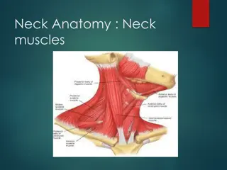
Anatomical Considerations Prominent landmarks Triangles of the neck Lymphatic levels Carotid bulb
Anatomical Considerations Prominent landmarks Triangles of the neck Lymphatic levels Carotid bulb
Anatomical Considerations Prominent landmarks Triangles of the neck Lymphatic levels Carotid bulb I
Anatomical Considerations Prominent landmarks Triangles of the neck Lymphatic levels Carotid bulb I
Anatomical Considerations Prominent landmarks Triangles of the neck Lymphatic levels Carotid bulb II I III IV
Anatomical Considerations Prominent landmarks Triangles of the neck Lymphatic levels Carotid bulb II I III IV
Anatomical Considerations Prominent landmarks Triangles of the neck Lymphatic levels Carotid bulb II I III V IV
Anatomical Considerations Prominent landmarks Triangles of the neck Lymphatic levels Carotid bulb
Anatomical Considerations Prominent landmarks Triangles of the neck Lymphatic levels Carotid bulb
General Considerations Patient age Pediatrics (0 15 years): mostly benign Young adults (16 40 years): similar to pediatric old adults (>40 years): High risk of malignancy Location Congenital masses: consistent in location Metastatic masses: key to primary lesion
Metastasis Location according to Various Primary Lesions
Diagnostic Steps History Developmental time course Associated symptoms (dysphagia, otalgia, voice) Personal habits (tobacco, alcohol) Previous irradiation or surgery Physical Examination Complete head and neck exam (visualize & palpate) Emphasis on location, mobility and consistency
Empirical Antibiotics Inflammatory mass suspected Two week trial of antibiotics Follow-up for further investigation
Diagnostic Tests Fine needle aspiration biopsy (FNAB) Computed tomography (CT) Magnetic resonance imaging (MRI) Ultrasonography Radionucleotide scanning
Fine Needle Aspiration Biopsy Standard of diagnosis Indications Any neck mass that is not an obvious abscess Persistence after a 2 week course of antibiotics Small gauge needle Reduces bleeding Seeding of tumor not a concern No contraindications (vascular ?)
Fine Needle Aspiration Biopsy Proper collection required Minimum of 4 separate passes Skilled cytopathologist essential On-site review best
Computed Tomography Distinguish cystic from solid Extent of lesion Vascularity (with contrast) Detection of unknown primary (metastatic) Pathologic node (lucent, >1.5cm, loss of shape) Avoid contrast in thyroid lesions
Magnetic Resonance Imaging Similar information as CT Better for upper neck and skull base Vascular delineation with infusion
Ultrasonography Less important now with FNAB Solid versus cystic masses Congenital cysts from solid nodes/tumors Noninvasive (pediatric)
Radionucleotide Scanning Salivary and thyroid masses Location glandular versus extra-glandular Functional information FNAB now preferred for for thyroid nodules Solitary nodules Multinodular goiter with new increasing nodule Hashimoto s with new nodule
Nodal Mass Workup in the Adult Any solid asymmetric mass MUST be considered a metastatic neoplastic lesion until proven otherwise Asymptomatic cervical mass 12% of cancer ~ 80% of these are SCCa
Nodal Mass Workup in the Adult Ipsilateral otalgia with normal otoscopy direct attention to tonsil, tongue base, supraglottis and hypopharynx Unilateral serous otitis direct examination of nasopharynx
Nodal Mass Workup in the Adult Panendoscopy FNAB positive with no primary on repeat exam FNAB equivocal/negative in high risk patient Directed Biopsy All suspicious mucosal lesions Areas of concern on CT/MRI None observed nasopharynx, tonsil (ipsilateral tonsillectomy for jugulodigastric nodes), base of tongue and piriforms Synchronous primaries (10 to 20%)
Nodal Mass Workup in the Adult Unknown primary University of Florida (August, 2001) Detected primary in 40% Without suggestive findings on CT or panendoscopy yield dropped to 20% Tonsillar fossa in 80%
Nodal Mass Workup in the Adult Open excisional biopsy Only if complete workup negative Occurs in ~5% of patients Be prepared for a complete neck dissection Frozen section results (complete node excision) Inflammatory or granulomatous culture Lymphoma or adenocarcinoma close wound
Primary Tumors Thyroid mass Lymphoma Salivary tumors Lipoma Carotid body and glomus tumors Neurogenic tumors
Thyroid Masses Leading cause of anterior neck masses Children Most common neoplastic condition Male predominance Higher incidence of malignancy Adults Female predominance Mostly benign
Thyroid Masses Lymph node metastasis Initial symptom in 15% of papillary carcinomas 40% with malignant nodules Histologically (microscopic) in >90% FNAB has replaced US and radionucleotide scanning Decreases # of patients with surgery Increased # of malignant tumors found at surgery Doubled the # of cases followed up Unsatisfactory aspirate repeat in 1 month
Lymphoma More common in children and young adults Up to 80% of children with Hodgkin s have a neck mass Signs and symptoms Lateral neck mass only (discrete, rubbery, nontender) Fever Hepatosplenomegaly Diffuse adenopathy
Lymphoma FNAB first line diagnostic test If suggestive of lymphoma open biopsy Full workup CT scans of chest, abdomen, head and neck; bone marrow biopsy
Salivary Gland Tumors Enlarging mass anterior/inferior to ear or at the mandible angle is suspect Benign Asymptomatic except for mass Malignant Rapid growth, skin fixation, cranial nerve palsies, pain
Salivary Gland Tumors Diagnostic tests Open excisional biopsy (submandibulectomy or parotidectomy) preferred FNAB Shown to reduce surgery by 1/3 in some studies Delineates intra-glandular lymph node, localized sialadenitis or benign lymphoepithelial cysts May facilitate surgical planning and patient counseling Accuracy >90% (sensitivity: ~90%; specificity: ~80%) CT/MRI deep lobe tumors, intra vs. extra-parotid Be prepared for total parotidectomy with possible facial nerve sacrifice
Carotid Body Tumor Rare in children Pulsatile, compressible mass Mobile medial/lateral not superior/inferior Clinical diagnosis, confirmed by angiogram or CT Treatment Irradiation or close observation in the elderly Surgical resection for small tumors in young patients Hypotensive anesthesia Preoperative measurement of catecholamines
Lipoma Soft, ill-defined mass Usually >35 years of age Asymptomatic Clinical diagnosis confirmed by excision
Neurogenic Tumors Arise from neural crest derivatives Include schwannoma, neurofibroma, and malignant peripheral nerve sheath tumor Increased incidence in NF syndromes Schwannoma most common in head & neck
Schwannoma Sporadic cases mostly 25 to 45% in neck when extracranial Most commonly between 20 and 50 years Usually mid-neck in poststyloid compartment Signs and symptoms Medial tonsillar displacement Hoarseness (vagus nerve) Horner s syndrome (sympathetic chain)
Congenital and Developmental Mass Epidermal and sebaceous cysts Branchial cleft cysts Thyroglossal duct cyst Vascular tumors
Epidermal and Sebaceous Cysts Most common congenital/developmental mass Older age groups Clinical diagnosis Elevation and movement of overlying skin Skin dimple or pore Excisional biopsy confirms
Branchial Cleft Cysts Branchial cleft anomalies 2nd cleft most common (95%) tract medial to XII nerve between internal and external carotids 1st cleft less common close association with facial nerve possible 3rd and 4th clefts rarely reported Present in older children or young adults often following URI
Branchial Cleft Cysts Most common as smooth, fluctuant mass underlying the SCM Skin erythema and tenderness if infected Treatment Initial control of infection Surgical excision, including tract May necessitate a total parotidectomy (1st cleft)
Thyroglossal Duct Cyst Most common congenital neck mass (70%) 50% present before age 20 Midline (75%) or near midline (25%) Usually just inferior to hyoid bone (65%) Elevates on swallowing/protrusion of tongue Treatment is surgical removal (Sis trunk) after resolution of any infection
Vascular Tumors Lymphangiomas and hemangiomas Usually within 1st year of life Hemangiomas often resolve spontaneously, while lymphangiomas remain unchanged CT/MRI may help define extent of disease
Vascular Tumors Treatment Lymphangioma surgical excision for easily accessible or lesions affecting vital functions; recurrence is common Hemangiomas surgical excision reserved for those with rapid growth involving vital structures or associated thrombocytopenia that fails medical therapy (steroids, interferon)
Inflammatory Disorders Lymphadenitis Granulomatous lymphadenitis
Lymphadenitis Very common, especially within 1st decade Tender node with signs of systemic infection Directed antibiotic therapy with follow-up FNAB indications (pediatric) Actively infectious condition with no response Progressively enlarging Solitary and asymmetric nodal mass Supraclavicular mass (60% malignancy) Persistent nodal mass without active infection