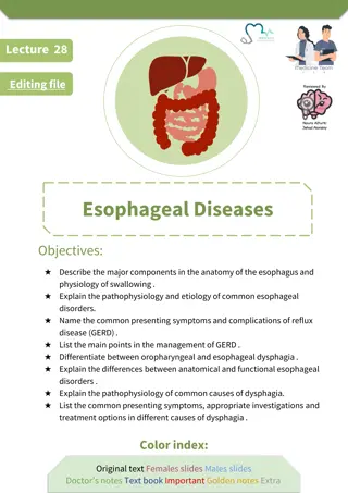
ESOPHAGEAL OBSTRUCTION
Esophageal obstruction can be acute or chronic, with various causes such as intraluminal or extraluminal obstructions, paralysis, strictures, and megaesophagus. Clinical signs include regurgitation, drooling, and bloat, impacting animals' ability to swallow.

Download Presentation

Please find below an Image/Link to download the presentation.
The content on the website is provided AS IS for your information and personal use only. It may not be sold, licensed, or shared on other websites without obtaining consent from the author.If you encounter any issues during the download, it is possible that the publisher has removed the file from their server.
You are allowed to download the files provided on this website for personal or commercial use, subject to the condition that they are used lawfully. All files are the property of their respective owners.
The content on the website is provided AS IS for your information and personal use only. It may not be sold, licensed, or shared on other websites without obtaining consent from the author.
E N D
Presentation Transcript
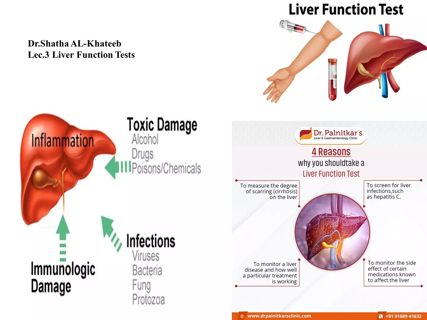
ESOPHAGEAL OBSTRUCTION By Dr. Hussein AlNaji
Esophageal obstruction can be acute or chronic and is characterized clinically by the inability to swallow, regurgitation of feed and water, continuous drooling of saliva, and bloat in ruminants. Acute cases are accompanied by signs of distress including retching and extension of the head. Horses with choke commonly regurgitate a mixture of saliva, feed, and water through the nostrils because of the anatomic characteristics of the equine soft palate.
ETIOLOGY Obstruction can be intraluminal and caused by swallowed material or extraluminal caused by pressure on the esophagus by surrounding organs or tissues. Esophageal paralysis can also result in obstruction, for example, in horses with grass sickness.
A.Intraluminal Obstructions 1. Solid obstructions, especially in cattle, by turnips, onions, potatoes, peaches, apples, oranges, and similar objects. 2. Fifteen-gram gelatin capsules in Shetland ponies. 3. Feedstuffs are a common cause of obstruction in horses and occasionally in other species. 4. Eating while sedated 5. Foreign bodies in horses include pieces of wood, antimicrobial boluses, and fragments of nasogastric tubes. 6. Atrichobezoar can cause esophageal obstruction cattle. 7. Poor dentition is often mooted.
B. Extraluminal Obstructions 1. Enlarged lymph nodes in the mediastinum (tuberculosis, neoplasia, Rhodococcus equi, Corynebacterium spp., strangles, and secondary to pleuritis). 2. Cervical or mediastinal abscess. 3. Persistent right aortic arch. 4. Thymoma 5. Megaesophagus and caudal esophageal muscle hypertrophy in Friesian horses can cause esophageal obstruction.
c. Esophageal Paralysis Esophageal paralysis can be caused by congenital or acquired abnormalities of the esophagus, and there are many examples of such abnormalities that interfere with swallowing and cause varying degrees of obstruction, even though it may be possible to pass a stomach tube through the esophagus into the stomach or rumen. D. Esophageal Strictures These arise as a result of cicatricial or granulation tissue deposition, usually as result of previous laceration or trauma of the esophagus.
E. Megaesophagus Megaesophagus is a dilatation and atony of the body of the esophagus usually associated with asynchronous function of the esophagus and the caudal esophageal sphincter. It occurs sporadically in cattle and in horses with preexisting esophageal disease. It is usually a congenital condition that causes regurgitation and aspiration pneumonia.
PATHOGENESIS An esophageal obstruction results in a physical inability to swallow and, in cattle, inability to eructate, with resulting bloat. In acute obstruction, there is initial spasm at the site of obstruction and forceful, painful peristalsis and swallowing movements. Complications of esophageal obstruction include 1. Laceration and rupture of the esophagus. 2. Esophagitis. 3. Stricture and stenosis. 4. The development of a diverticulum.
Acquired esophageal diverticula can occur in the horse. A traction diverticulum occurs following periesophageal scarring and is of little consequence. An esophageal pulsion diverticulum is a circumscribed sac of mucosa protruding through a defect in the muscular layer of the esophagus.
Complications associated with esophageal diverticula include 1. Peridiverticulitis, 2. Pulmonary adhesions. 3. Abscesses, and mediastinitis. In megaesophagus, the esophagus is dysfunctional, dilated, and filled with saliva, feed, and water. This results in regurgitation and can lead to aspiration pneumonia.
CLINICAL FINDINGS Acute Obstruction or Choke Cattle The obstruction is usually in the cervical esophagus just above the larynx or at the thoracic inlet. Obstructions can also occur at the base of the heart or the cardia. 1. The animal suddenly stops eating and shows anxiety and restlessness. 2. There are forceful attempts to swallow and regurgitate, salivation, coughing, and continuous chewing movements.
3- If obstruction is complete, bloating occurs rapidly and adds to the animal s discomfort. 4- Ruminal movements are continuous and forceful and there can be a systolic murmur audible on auscultation of the heart. 5- The acute signs, other than bloat, usually disappear within a few hours. This is caused by relaxation of the initial esophageal spasm and can or cannot be accompanied by onward passage of the obstruction.
Horse In the horse with esophageal obstruction caused by feed, the obstruction can occur at any level of the esophagus from the upper cervical region all the way to the thoracic portion. 1. Dysphagia with nasal reflux of saliva, feed, and water. 2. Affected horses will usually not attempt further eating but will drink and attempt to swallow water. 3. External palpation of the cervical esophagus can reveal a firm cylindrical swelling along the course of the neck on the left side when the esophagus is obstructed with feed.
4- Horses with acute esophageal obstruction are commonly difficult to handle because they are panicky and make forceful attempts to swallow or retch. 5- They often vigorously extend and flex their necks and stamp their front feet. Chronic Obstruction No acute signs of obstruction are evident and in cattle the earliest sign is chronic bloat, which is usually of moderate severity and can persist for several days without the appearance of other signs.
DIFFERENTIAL DIAGNOSIS 1. The clinical findings of acute esophageal obstruction in cattle and horses are usually typical but can be similar to those of esophagitis, in which local pain is more apparent and there is often an accompanying stomatitis and pharyngitis. 2. The excitement, sweating, and tachycardia observed in acute choke in the horse often suggests colic. Passage of a nasogastric tube reveals the obstruction. The use of a fiberoptic endoscope will usually locate the obstruction for visualization, and obstructions are easiest to see when the endoscope is being withdrawn rather than advanced.
Chronic Obstruction 1. Mediastinal lymph node enlargement is usually accompanied by other signs of tuberculosis or lymphomatosis. 2. Chronic ruminal tympany in cattle can be caused by ruminal atony, in which case there is an absence of normal ruminal movements. 3. Diaphragmatic hernia can also be a cause of chronic ruminal tympany in cattle and is sometimes accompanied by obstruction of the esophagus with incompletely regurgitated ingesta. This condition and vagus indigestion, another cause of chronic tympany, are usually accompanied by a systolic cardiac murmur, but passage of a stomach tube is unimpeded. 4. Equine encephalomyelitis and botulism are other diseases in which there is difficulty in swallowing. 5. Cleft palate is a cause of recurrent nasal regurgitation in foals.
TREATMENT a. ConservativeApproach Many obstructions will resolve spontaneously and a careful conservative approach is recommended. a. Sedation In acute obstruction, if there is marked anxiety and distress, the animal should be sedated before proceeding with specific treatment. For sedation and esophageal relaxation in the horse, one of the following is recommended: 1. Acepromazine 0.05 mg/kg BW intravenously 2. Xylazine 0.5 to 1.0 mg/kg BW intravenously 3. Detomidine 0.01 to 0.02 mg/kg BW intravenously. 4. Romifidine 0.04 to 0.12 mg/kg intravenously.
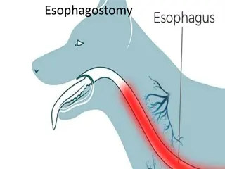
For esophageal relaxation, analgesia and anti-inflammatory effect hyoscine: dipyrone 0.5 : 0.22 mg/kg BW intravenously for analgesia and anti-inflammatory effect flunixin meglumine 1.1 mg/kg BW intravenously or phenylbutazone 2 to 4 mg/ kg intravenously are suggested. a. Pass a Stomach Tube andAllow Object to Move Into Stomach. b. Manual Removal Through Oral Cavity in Cattle. c. Esophageal Lavage in the Horse. d. Surgical Removal of Foreign Bodies. e. Cervical EsophagostomyAlimentation. f. AntimicrobialAdministration.