Endophthalmitis: Causes, Risks, and Management
 undefined


PROF. SANDEEP SAXENA
MS,FRCS(ED),FRCS
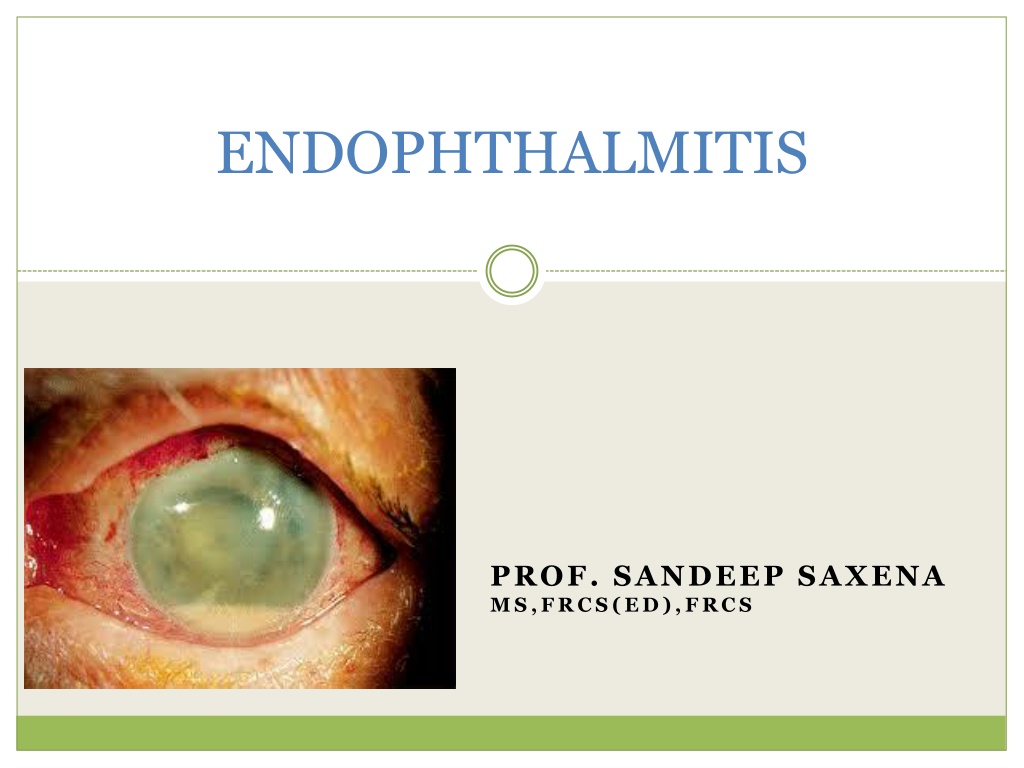
ENDOPHTHALMITIS
DEFINITION
An intraocular
inflammation
involving ocular
cavities (vitreous
cavity and/ or anterior
chamber) and their
adjacent structures
.
CLASSIFICATION
INFECTIOUS
Exogenous
-Acute onset -Post traumatic
-Delayed onset
-Bleb associated
Endogenous
-Haematogenous spread
STERILE
Lens induced
Toxic
Post surgical
Non surgical
CAUSATIVE ORGANISMS
Gram +ve: Bacteria: Bacteria:
Bacteria:
S. epidermidis Propionibacterium Bacillus Bacillus cereus
S. aureus acne S.epidermidis Streptococci
Streptococci Streptococci Streptococci S.aureus
Gram –ve: Fungi: Fungi:
N.meningitides
Pseudomonas Aspergillus Fusarium H.influenzae
H.influenzae Candida
Fungi:
Klebsiella spp Fusarium Mucor
E. coli Penicillium Candida
Bacillus spp
Anaerobes
Acute Post-op
Delayed Post-op
Post- Traumatic
Endogenous
POST- SURGICAL ENDOPHTHALMITIS
Most common form
70% cases of infective endophthalmitis
Worldwide incidence 0.04 - 4%
Incidence in India 0.7 - 2.4%
Commonly associated with:
Cataract extraction (most common)
Secondary lens implantation
Pars plana vitrectomy
Glaucoma filteration surgery
Penetrating keratoplasty
RISK FACTORS
PRE- OPERATIVE RISK FACTORS:
Blepharitis
Conjunctivitis
Lacrimal drainage system infection
Contact lenses wear
Contaminated eyedrops
INTRA-OP RISK FACTORS:
Clear corneal incision
Temporal incision
Posterior capsule rupture
Vitreous incarceration in wound
Prolonged procedure time
Contaminated irrigation solutions
Combined procedures
POST- OPERATIVE RISK FACTORS:
Inadequately buried sutures
Wound leak on the first day
Delaying post-op topical antibiotics until the day after
surgery
CLINICAL PRESENTATION
Acute onset
Delayed onset
Within 6 weeks
After 6 weeks
ACUTE POST-OP ENDOPHTHALMITIS
Most common organism - Coagulase negative
Staphylococcus
species (
S.epidermidis)
Hyperacute infections -
Pseudomonas aeruginosa
and
Bacillus
species.
Source of infection- lid flora
- conjunctival flora
Entry occurs at the time of surgery
DELAYED- ONSET ENDOPHTHALMITIS
Low virulence organisms:
•
Propionibacterium acne
•
Staphylococcus epidermidis
•
Fungi
Release of organisms sequestered within the
capsular bag- saccular endophthalmitis
CLINICAL FEATURES
SYMPTOMS:
Blurred vision (94%)
Red eye (82%)
Pain (74%)
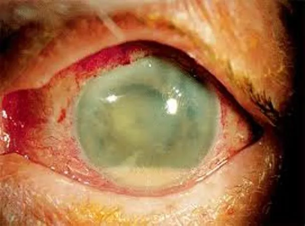
CLINICAL FEATURES
SIGNS:
Decreased visual acuity
Lid edema, conjunctival chemosis, congestion and discharge
Corneal edema
Keratic precipitates (delayed-onset)
Fibrinous exudates and hypopyon in AC
SIGNS
Relative afferent pupillary defect
Loss of red reflex, impaired fundal view, vitritis
Scattered retinal haemorrhages, periphlebitis
Capsular plaque (Propionibacterium acnes endophthalmitis)
BLEB- ASSOCIATED ENDOPHTHALMITIS
Incidence:
•
Acute- 0.06-0.2%
Predisposing factors:
Blepharitis
Use of anti- fibrotic agents (Mitomycin- C, 5- fluorouracil
)
Long term topical antibiotic use
Inferior or nasally placed bleb
Bleb leak
Pathogens:
Streptococcus
H.influenzae
Staphylococcus
•
Delayed- 0.2-18%
POST- TRAUMATIC ENDOPHTHALMITIS
Occurs following penetrating trauma (7%)
Intraocular foreign body increases the risk (30%)
Common organisms inolved:
•
Gram positive cocci
•
Bacillus spp
•
Fungi (esp. Fusarium)
May occur anytime from days to weeks following
injury
Delay in diagnosis: Post- traumatic inflammation vs
infection
ENDOGENOUS ENDOPHTHALMITIS
Haematogenous spread of micro-organisms from a site
external to the eye
Predisposing host factors:
Age (children)
Immune suppression
Malnutrition
Diabetes mellitus
Alcoholism
Malignancy
Presents with less inflammation and pain than other forms
of endophthalmitis
Reduced vision and floaters in one or both eyes
DIAGNOSIS OF ENDOPHTHALMITIS
Early recognition is critical.
High index of suspicion to be maintained.
A complete ocular and medical history is essential.
Thorough ophthalmic examination performed.
OPHTHALMIC INVESTIGATIONS
Conjunctival swab
For pre-existing organisms in adnexae
Ultrasonography
Useful in anterior segment media opacity
Confirm presence of variable echoes in vitreous
Retained lens remnants in posterior segment
Intraocular foreign body in post- traumatic cases
Retinal or choroidal detachment
Provide a baseline to compare
IDENTIFICATION OF PATHOGENS
Aqueous tap:
0.1-0.2 ml of aqueous is aspirated via a limbal paracentesis
using a 25-G needle
Vitreous tap:
0.2-0.4 ml is aspirated from mid-vitreous cavity using a 23-G
needle
Distance from limbus-
3mm for aphakic eye
3.5mm for pseudophakic eye
4mm for phakic eye
Samples are subjected to:
Gram staining
Giemsa staining
KOH mount
Culture on
-
Blood agar
Chocolate agar
Sabouraud dextrose agar
Thioglycollate broth
Anaerobic medium
Polymerase chain reaction
Reasons for negative culture results:
Fastidious organisms
Insufficient sampling
Sterile endophthalmitis
Repeat cultures may be needed:
When clinical response is not good
Presence of contaminants in media
Presence of fungus- especially likely to be missed initially
SYSTEMIC INVESTIGATIONS
Complete haemogram
Blood sugar (predisposition in diabetics)
Blood and urine cultures (endogenous endophthalmitis)
Cultures from other sites (catheter tips, skin wounds,
abscesses and joints)
TREATMENT
Antibiotics
Steroids
Topical mydriatics
Vitrectomy
IOL management
Evisceration
MEDICAL
SURGICAL
INTRAVITREAL ANTIBIOTICS
Gram positive:
Vancomycin (1.0 mg in 0.1 ml)
Broad spectrum
B
oth coagulase positive and coagulase negative cocci
Gram negative:
Ceftazidime (2.25 mg in 0.1 ml)
No retinal toxicity
Amikacin (0.4 mg in 0.1 ml)
Retinotoxic
Alternative to ceftazidime in penicillin allergy
Gentamicin
OTHER MODES
Topical antibiotics:
Fortified cefazoline (5%) OR Fortified vancomycin (5%)
PLUS
Fortified tobramycin (1.3%)
Given half hourly alternately
Systemic antibiotics:
Clindamycin 1g iv 8 hrly
Ceftazidime 2g iv 8 hrly
Ciprofloxacin 750 mg P.O. bid
Moxifloxacin 400 mg P.O. od
STEROIDS
Control inflammation mediated damage
But no influence on visual outcome
INTRAVITREAL:
Dexamethasone (0.4 mg in 0.1 ml)
Triamcinolone (long acting) can also be used
SUBCONJUNCTIVAL:
Dexamethasone (6mg in 0.25 ml)
TOPICAL:
Prednisolone 1% 2 hrly
Dexamethasone 0.1%
SYSTEMIC:
Prednisolone 1mg/kg OD (started after 12-24 hrs)
FUNGAL INFECTION
Intravitreal Amphotericin B (5µg in 0.1 ml)
Newer agents- Voriconazole (200µg in 0.1 ml) and
Caspofungin
Topical Natamycin (5%) and Itraconazole (1%)
Systemic therapy- Fluconazole (150mg od)
Steroids are contraindicated
SURGICAL MANAGEMENT
VITRECTOMY:
Advantages of early vitrectomy:
Clearing of ocular media
Reduction of bacterial load
Removal of bacterial products
Removal of vitreous scaffolding- which may cause retinal
detachment
Disadvantages:
Iatrogenic retinal holes and detachments
Choroidal haemorrhage
Retinal detachment - difficult to treat in vitrectomized eyes
COMPLICATIONS RELATED TO IOL
Fibrin exudates on IOL- removed with a needle or forceps
Exudates trapped between the posterior capsule and IOL -
Posterior capsulotomy
Fungal endophthalmitis and sequestered organisms in the
capsular bag (P.acnes) - en bloc removal of IOL and
capsular bag
MANAGEMENT PROTOCOL
Assess
visual
acuity
Only PL+
Early VIT
+ i/vit
Antibiotics
HM or
better
I/vit
Antibiotics
Improves
Does not
improve
•
Repeat
i/vit
antibiotics
•
Vitrectomy
•
Repeat
culture
Watch
for 48
hours
EMPIRICAL MEDICAL THERAPY OF
ENDOPHTHALMITIS
(as per EVS 1996)
ACUTE ONSET POST CATARACT EXTRACTION
INTRAVITREAL
Vancomycin
Ceftazidime OR amikacin
Dexamethasone (optional)
SUBCONJUNCTIVAL
Vancomycin
Ceftazidime or Amikacin (if B-lactam allergy)
Dexamethasone
TOPICAL
Vancomycin hydrochloride
Amikacin
Atropine sulphate
Prednisolone acetate 1%
ORAL
Prednisone 30mg twice daily for 5 to 10 days (optional)
POST- TRAUMATIC
Same as that for Post- cataract Sx with:
Intravitreal Clindamycin (450 micrograms)
Systemic antibiotics
BLEBITIS
Topicals are sufficient:
Vancomycin hydrochloride
Amikacin
Atropine sulphate
Prednisolone acetate 1%
BLEB- ASSOCIATED ENDOPHTHALMITIS
Same as that for Post- cataract Sx with systemic antibiotics
FOLLOW-UP AND OUTCOME
Signs of improvement:
AC reaction
Hypopyon
Fundal glow
Final outcome:
Duration of infection
Virulence of organism
(EVS- Final outcomes)
53% patients visual acuity >6/12
75% patients visual acuity >6/30
89% patients visual acuity >6/240
PROPHYLAXIS
5% povidone iodine - 3 minutes
Treatment of pre-existing infections
Prophylactic antibiotics:
Pre-operative topical fluoroquinolones
Intracameral cefuroxime (1 mg in 0.1 ml)
Post-operative sub-conjunctival antibiotics
Systemic 4
th
generation fluoroquinolones
Early resuturing of wound leaks
Endophthalmitis is an intraocular inflammation that can result from various causes, including post-surgical procedures and infections. It can lead to serious complications if not managed promptly. Understanding the classification, causative organisms, associated risk factors, and clinical presentation is essential for effective treatment and prevention.

Download Presentation

Please find below an Image/Link to download the presentation.
The content on the website is provided AS IS for your information and personal use only. It may not be sold, licensed, or shared on other websites without obtaining consent from the author.If you encounter any issues during the download, it is possible that the publisher has removed the file from their server.
You are allowed to download the files provided on this website for personal or commercial use, subject to the condition that they are used lawfully. All files are the property of their respective owners.
The content on the website is provided AS IS for your information and personal use only. It may not be sold, licensed, or shared on other websites without obtaining consent from the author.
E N D
Presentation Transcript
ENDOPHTHALMITIS PROF. SANDEEP SAXENA MS,FRCS(ED),FRCS
DEFINITION An intraocular inflammation involving ocular cavities (vitreous cavity and/ or anterior chamber) and their adjacent structures.
CLASSIFICATION INFECTIOUS Exogenous STERILE Lens induced Toxic Post surgical Non surgical -Acute onset -Post traumatic -Delayed onset -Bleb associated Endogenous -Haematogenous spread
CAUSATIVE ORGANISMS Acute Post-op Delayed Post-op Post- Traumatic Endogenous Gram +ve: Bacteria: Bacteria: S. epidermidis Propionibacterium Bacillus Bacillus cereus S. aureus acne S.epidermidis Streptococci Streptococci Streptococci Streptococci S.aureus Gram ve: Fungi: Fungi: N.meningitides Pseudomonas Aspergillus Fusarium H.influenzae H.influenzae Candida Fungi: Klebsiella spp Fusarium Mucor E. coli Penicillium Candida Bacillus spp Anaerobes Bacteria:
POST- SURGICAL ENDOPHTHALMITIS Most common form 70% cases of infective endophthalmitis Worldwide incidence 0.04 - 4% Incidence in India 0.7 - 2.4%
Commonly associated with: Cataract extraction (most common) Secondary lens implantation Pars plana vitrectomy Glaucoma filteration surgery Penetrating keratoplasty
RISK FACTORS PRE- OPERATIVE RISK FACTORS: Blepharitis Conjunctivitis Lacrimal drainage system infection Contact lenses wear Contaminated eyedrops
INTRA-OP RISK FACTORS: Clear corneal incision Temporal incision Posterior capsule rupture Vitreous incarceration in wound Prolonged procedure time Contaminated irrigation solutions Combined procedures
POST- OPERATIVE RISK FACTORS: Inadequately buried sutures Wound leak on the first day Delaying post-op topical antibiotics until the day after surgery
CLINICAL PRESENTATION Acute onset Delayed onset Within 6 weeks After 6 weeks
ACUTE POST-OP ENDOPHTHALMITIS Most common organism - Coagulase negative Staphylococcus species (S.epidermidis) Hyperacute infections - Pseudomonas aeruginosa and Bacillus species. Source of infection- lid flora - conjunctival flora Entry occurs at the time of surgery
DELAYED- ONSET ENDOPHTHALMITIS Low virulence organisms: Propionibacterium acne Staphylococcus epidermidis Fungi Release of organisms sequestered within the capsular bag- saccular endophthalmitis
CLINICAL FEATURES SYMPTOMS: Blurred vision (94%) Red eye (82%) Pain (74%)
CLINICAL FEATURES SIGNS: Decreased visual acuity Lid edema, conjunctival chemosis, congestion and discharge Corneal edema Keratic precipitates (delayed-onset) Fibrinous exudates and hypopyon in AC
SIGNS Relative afferent pupillary defect Loss of red reflex, impaired fundal view, vitritis Scattered retinal haemorrhages, periphlebitis Capsular plaque (Propionibacterium acnes endophthalmitis)
BLEB- ASSOCIATED ENDOPHTHALMITIS Incidence: Acute- 0.06-0.2% Predisposing factors: Blepharitis Use of anti- fibrotic agents (Mitomycin- C, 5- fluorouracil) Long term topical antibiotic use Inferior or nasally placed bleb Bleb leak Delayed- 0.2-18% Pathogens: Streptococcus H.influenzae Staphylococcus
POST- TRAUMATIC ENDOPHTHALMITIS Occurs following penetrating trauma (7%) Intraocular foreign body increases the risk (30%) Common organisms inolved: Gram positive cocci Bacillus spp Fungi (esp. Fusarium) May occur anytime from days to weeks following injury Delay in diagnosis: Post- traumatic inflammation vs infection
ENDOGENOUS ENDOPHTHALMITIS Haematogenous spread of micro-organisms from a site external to the eye Predisposing host factors: Age (children) Immune suppression Malnutrition Diabetes mellitus Alcoholism Malignancy Presents with less inflammation and pain than other forms of endophthalmitis Reduced vision and floaters in one or both eyes
DIAGNOSIS OF ENDOPHTHALMITIS Early recognition is critical. High index of suspicion to be maintained. A complete ocular and medical history is essential. Thorough ophthalmic examination performed.
OPHTHALMIC INVESTIGATIONS Conjunctival swab For pre-existing organisms in adnexae Ultrasonography Useful in anterior segment media opacity Confirm presence of variable echoes in vitreous Retained lens remnants in posterior segment Intraocular foreign body in post- traumatic cases Retinal or choroidal detachment Provide a baseline to compare
IDENTIFICATION OF PATHOGENS Aqueous tap: 0.1-0.2 ml of aqueous is aspirated via a limbal paracentesis using a 25-G needle Vitreous tap: 0.2-0.4 ml is aspirated from mid-vitreous cavity using a 23-G needle Distance from limbus- 3mm for aphakic eye 3.5mm for pseudophakic eye 4mm for phakic eye
Samples are subjected to: Gram staining Giemsa staining KOH mount Culture on- Blood agar Chocolate agar Sabouraud dextrose agar Thioglycollate broth Anaerobic medium Polymerase chain reaction
Reasons for negative culture results: Fastidious organisms Insufficient sampling Sterile endophthalmitis Repeat cultures may be needed: When clinical response is not good Presence of contaminants in media Presence of fungus- especially likely to be missed initially
SYSTEMIC INVESTIGATIONS Complete haemogram Blood sugar (predisposition in diabetics) Blood and urine cultures (endogenous endophthalmitis) Cultures from other sites (catheter tips, skin wounds, abscesses and joints)
TREATMENT MEDICAL SURGICAL Antibiotics Steroids Topical mydriatics Vitrectomy IOL management Evisceration
INTRAVITREAL ANTIBIOTICS Gram positive: Vancomycin (1.0 mg in 0.1 ml) Broad spectrum Both coagulase positive and coagulase negative cocci Gram negative: Ceftazidime (2.25 mg in 0.1 ml) No retinal toxicity Amikacin (0.4 mg in 0.1 ml) Retinotoxic Alternative to ceftazidime in penicillin allergy Gentamicin
OTHER MODES Topical antibiotics: Fortified cefazoline (5%) OR Fortified vancomycin (5%) PLUS Fortified tobramycin (1.3%) Given half hourly alternately Systemic antibiotics: Clindamycin 1g iv 8 hrly Ceftazidime 2g iv 8 hrly Ciprofloxacin 750 mg P.O. bid Moxifloxacin 400 mg P.O. od
STEROIDS Control inflammation mediated damage But no influence on visual outcome INTRAVITREAL: Dexamethasone (0.4 mg in 0.1 ml) Triamcinolone (long acting) can also be used SUBCONJUNCTIVAL: Dexamethasone (6mg in 0.25 ml) TOPICAL: Prednisolone 1% 2 hrly Dexamethasone 0.1% SYSTEMIC: Prednisolone 1mg/kg OD (started after 12-24 hrs)
FUNGAL INFECTION Intravitreal Amphotericin B (5 g in 0.1 ml) Newer agents- Voriconazole (200 g in 0.1 ml) and Caspofungin Topical Natamycin (5%) and Itraconazole (1%) Systemic therapy- Fluconazole (150mg od) Steroids are contraindicated
SURGICAL MANAGEMENT VITRECTOMY: Advantages of early vitrectomy: Clearing of ocular media Reduction of bacterial load Removal of bacterial products Removal of vitreous scaffolding- which may cause retinal detachment
Disadvantages: Iatrogenic retinal holes and detachments Choroidal haemorrhage Retinal detachment - difficult to treat in vitrectomized eyes
COMPLICATIONS RELATED TO IOL Fibrin exudates on IOL- removed with a needle or forceps Exudates trapped between the posterior capsule and IOL - Posterior capsulotomy Fungal endophthalmitis and sequestered organisms in the capsular bag (P.acnes) - en bloc removal of IOL and capsular bag
MANAGEMENT PROTOCOL Early VIT + i/vit Antibiotics Only PL+ Assess visual acuity Improves Watch for 48 hours Repeat i/vit antibiotics Vitrectomy Repeat culture HM or better I/vit Antibiotics Does not improve
EMPIRICAL MEDICAL THERAPY OF ENDOPHTHALMITIS (as per EVS 1996) ACUTE ONSET POST CATARACT EXTRACTION INTRAVITREAL Vancomycin Ceftazidime OR amikacin Dexamethasone (optional) SUBCONJUNCTIVAL Vancomycin Ceftazidime or Amikacin (if B-lactam allergy) Dexamethasone
TOPICAL Vancomycin hydrochloride Amikacin Atropine sulphate Prednisolone acetate 1% ORAL Prednisone 30mg twice daily for 5 to 10 days (optional)
POST- TRAUMATIC Same as that for Post- cataract Sx with: Intravitreal Clindamycin (450 micrograms) Systemic antibiotics BLEBITIS Topicals are sufficient: Vancomycin hydrochloride Amikacin Atropine sulphate Prednisolone acetate 1% BLEB- ASSOCIATED ENDOPHTHALMITIS Same as that for Post- cataract Sx with systemic antibiotics
FOLLOW-UP AND OUTCOME Signs of improvement: AC reaction Hypopyon Fundal glow Final outcome: Duration of infection Virulence of organism (EVS- Final outcomes) 53% patients visual acuity >6/12 75% patients visual acuity >6/30 89% patients visual acuity >6/240
PROPHYLAXIS 5% povidone iodine - 3 minutes Treatment of pre-existing infections Prophylactic antibiotics: Pre-operative topical fluoroquinolones Intracameral cefuroxime (1 mg in 0.1 ml) Post-operative sub-conjunctival antibiotics Systemic 4th generation fluoroquinolones Early resuturing of wound leaks