Differential Leucocyte Count for Blood Disorders
Differential Leucocyte Count (DLC) is crucial for diagnosing blood-related disorders by analyzing the distribution and morphology of white blood cells. Dr. Versha Prasad explains the significance, normal values, and various conditions like neutrophilia, lymphocytosis, monocytosis, and basophilia. The procedure requires a blood specimen in EDTA anticoagulated blood. Learn about the requirements, specimen preparation, and staining techniques for accurate results.

Download Presentation

Please find below an Image/Link to download the presentation.
The content on the website is provided AS IS for your information and personal use only. It may not be sold, licensed, or shared on other websites without obtaining consent from the author.If you encounter any issues during the download, it is possible that the publisher has removed the file from their server.
You are allowed to download the files provided on this website for personal or commercial use, subject to the condition that they are used lawfully. All files are the property of their respective owners.
The content on the website is provided AS IS for your information and personal use only. It may not be sold, licensed, or shared on other websites without obtaining consent from the author.
E N D
Presentation Transcript
Differential Leucocyte Count Dr Versha Prasad Dr Versha Prasad
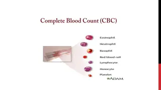
Introduction DLC is important for the diagnosis of various blood related disorders, involving white cell or red cell. Generally, it is performed to check the normal number or distribution of different leucocytes. It also gives morphology of different cells.It is usefull to identify changes in the distribution of white cells which may be related to specific types of disorders. Dr Versha Prasad
Neutrophilia Increase in % of neutrophil is called neutrophilia. The commonest cause is pyogenic infection. NEUTROPENIA Decrease in % of neutrophil is observed in infections such as bacterial, viral and in other conditions such as anemia and in supression of bone marrow. Dr Versha Prasad
Lymphocytosis Increase in Lymphocytes. Occurs in infections such as mumps, measles, influenza, syphilis, T.B. ,Typhoid. Eosinophilia- Increase in eosinophil count. Seen in allergic conditions and hypersensitivity reaction and in parasitic infections. Dr Versha Prasad
Monocytosis- Increase in monocte % .seen in T.B. malaria Typhoid. Basophilia- Observed in CML ( Chronic Myeloid Leukemia). Dr Versha Prasad
Normal Values Cells In % Per cu mm Neutrophil 60 to 70 3000 to 7000 Lymphocyte 25 to 35 2000 to 3000 Monocyte 2 to 6 100 to 600 Eosinophil 1 to 6 50 to 400 Basophil 0 to o.5 0 to 50 Thus DLC gives us the idea about distribution of different proportion of white cells Dr Versha Prasad
Specimen The blood specimen to be used for differential leucocytes count is EDTA anticoagulated blood. The blood smear should be preferably prepared immediately after skin puncture or Venipunture before mixing the anticoagulant. If EDTA blood is used then smear should be prepared with in 1-2 hrs after blood withdrawal. Dr Versha Prasad
Requirements Sterilized Needle. Cotton. Sprit 2 perfectly grease free dried slides with smooth edges. Dr Versha Prasad
Leishmans stain- Prepared by Dissolving 0.15 G of powdered stain in 100 ml of acetone free Methyl Alcohol. This is a polychromic staining solutions contain metylene blue and eosin. These acidic and basic dyes induces multiple colour when applied to cells. Methanol act as a fixative and also as a solvent. The fixative does not allow any further changes in the cells and make them adhere to the glass slide. The basic components of WBC i.e. cytoplasm is stained by acidic dye and they are described as eosinophilic or acidophilic. The acidic components ( e.g. nucleus) take blue to purple shades by the basic dye and they are called basophilic. The neutral components of the cells are stained by both the dyes. Dr Versha Prasad
Blood cells have different structures, which takes different stains. Some are basophilic, others are acidophilic while some cells accepts neutral stain. Therefore stain used in combination of these three. Such stains are called as Romanowsky Stain . The three different types of Romanowsky Stain are commonly in use. i) Leishman stain ii) Giemsa s stain iii) Wright s Stain. Dr Versha Prasad
Procedure Take aclean, dry slide. Transfer a small drop of blood near the edge of the slide. Place the spreader slide at an angle of 30 . Pull back the spreader untill it touches the drop of blood. Let the blood run along the edge of the spreader. Push the spreader forward to the end of the slide with a smooth movement. Dry the smear at room temperature. Drying is essential to preserve the quality of the film. 1. 2. 3. 4. 5. Dr Versha Prasad
Precautions Dirty slides do not give an even smear. Use an appropriate size of blood drop. After putting the drop on the slide, make the smear immediately for even distribution of white cells on the slide. The thickness of smear depends on the angle of the spreader. If the angle is less than 30 - Thinner smear. If more than 30 - thicker smear. The film must be smooth at the end. There should be no lines extending across or down through the film and it should not contain holes. 1. 2. 3. 4. 5. Dr Versha Prasad
Criteria of a good thin film a) Surface: even and uniform; free from ridges, waves and holes. b) Margin; will not touch the sides of the slide. Tail end: will be gradually thin. Without any serrated end. Ends near about the center of the slide c) Dr Versha Prasad
d) In an ideal smear it consists three parts: Head, Body and Tail. e) Length: about 2.5 cm long. Dr Versha Prasad
Identification Marking By using lead pencil or marker pen always write identification no. on the slide. Fixing the smear. - - The slide should be stained after making the smear. Methanol present in the stain fixes the smear. Dr Versha Prasad


 In an ideal smear it consists three")