Cholecystitis and Cholelithiasis: Causes, Risk Factors, and Management
Inflammation of the gallbladder (cholecystitis) and the presence of stones in the gallbladder (cholelithiasis) are common conditions. Acute cholecystitis is often caused by gallstones, while chronic cholecystitis can result from repeated attacks or chronic irritation. Risk factors include gender, age, obesity, and lifestyle choices. Cholelithiasis is more prevalent in women, especially above 40 and obese individuals. Understanding the causes and risk factors is crucial for prevention and management.

Download Presentation

Please find below an Image/Link to download the presentation.
The content on the website is provided AS IS for your information and personal use only. It may not be sold, licensed, or shared on other websites without obtaining consent from the author.If you encounter any issues during the download, it is possible that the publisher has removed the file from their server.
You are allowed to download the files provided on this website for personal or commercial use, subject to the condition that they are used lawfully. All files are the property of their respective owners.
The content on the website is provided AS IS for your information and personal use only. It may not be sold, licensed, or shared on other websites without obtaining consent from the author.
E N D
Presentation Transcript
Lecture #5 First semester Lecture #7 cholecystitis and cholelithiasis By : :by Assistant Prof. Dr. Raad A. AL-Harmoosh Professor Dr. Fakhria Jaber Al-Toosi University College Department of Medical Laboratory Techniques 4thClass Diagnostic Microbiology Department of Nursing Al-Mustaqbel University College 2nd Class Adult Nursing
Cholecystitis Definition: It is inflammation of the gallbladder (may be acute or chronic). 1. Acute cholecystitis is an acute infection of the gallbladder . calculus cholecystitis most cases are caused by gallstone obstruction of the cystic duct, causing edema, inflammation, and bacterial invasion. Thanks For Listening A calculus cholecystitis is acute gallbladder inflammation in the absence of obstruction by gallstones. 2. Chronic cholecystitis occurs when the gallbladder becomes thickened, rigid, and fibrotic and functions poorly . Results from repeated attacks of cholecystitis, presence of calculi, or chronic irritation.
Incidence & Risk Factors Acute cholecystitis can affect any age group. Chronic cholecystitis primarily affects middle aged and obese older women. Incidence of chronic cholecystitis has a female to male ratio of 4:1. A higher incidence of cholecystitis is seen among individuals with a sedentary life style.
In acute cholecystitis the risk factors are following: 1. Gall stone. 2. Sepsis due to invasion by bacteria (usually Escherichia coli, salmonella or streptococcus) via lymphatic or vascular route. 3. Multiple child birth. 4. Prolonged dehydration. 5. Prolonged immobility. In chronic cholecystitis the risk factors are following: 1. Cholelithiasis. 2. Obesity. 3. Acute cholecystitis
Cholelithiasis Definition: Is the presence of stones in the gallbladder. Gallstone formation takes place in the gallbladder, the common bile duct, and the duodenum. Types of gallstones: a.Cholesterol gallstones contain at least 50% cholesterol by weight. b.Pigmented gallstones are made up of bile pigments and are caused by increased production and execration of bilirubin or stasis of bile such as cirrhosis, hemolysis, and infection of the biliary tree.
Risk Factors: a.Women are 4 times risk more than men to develop cholesterol stones, usually above 40 & obese, (4 F's: female, fatty, fertile, forty). a.The incidence of stone formation rises in users of oral contraceptives, estrogen. b.The incidence increases with age as a result of increased hepatic secretion of cholesterol and decreased bile acid synthesis. c. People with diabetes.
Pathophysiology Stones occur when cholesterol supersaturates the bile in the gallbladder and precipitates out of the bile. The cholesterol-saturated bile predisposes to the formation of gallstones and acts as an irritant, producing inflammatory changes in the gallbladder .
Clinical Manifestations 1. Biliary colic can be caused by the presence of gallstones . a) Steady, severe aching pain or sensation of pressure in the epigastrium or right upper quadrant, which may radiate to the right scapular area or right shoulder. b) Epigastric distress as fullness and abdominal distention epically after a fatty meal. c) Begins suddenly and persists for 1 to 3 hours until the stone falls back into the gallbladder or is passed through the cystic duct . 2. Acute cholecystitis causes biliary colic pain that persists more than 4 hours and increases with movement, including respirations .
a) Also causes nausea and vomiting, low-grade fever, and jaundice (with stones or inflammation in the common bile duct .( b) Right upper quadrant guarding and Murphy s sign (inability to take a deep inspiration when examiner s fingers are pressed below the hepatic margin) are present . 3 - Chronic cholecystitis causes a. Heartburn, flatulence, and eructation because of gas accumulation resulting from impaired fat digestion b.Jaundice from obstruction of the common bile duct, the bile is absorbed by the blood and gives the skin and mucous membrane a yellow color causing marked itching. c. Clay colored stools. d. Steattorrhea ( presence of fat in the stool)
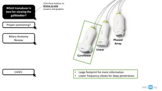
Diagnostic Evaluation 1.Oral cholecystography, ultrasonography, may visualize stones or inflammation . 2.ERCP (Endoscopic retrograde Cholangiopancreatography)to visualize location of stones and obstruction . 3.Elevated conjugated bilirubin due to obstruction .
Clinical Management B- Non Pharmacological interventions: 1.A low fat diet to decrease stimulation of the gall bladder. 2.Weight reduction, including prescribed diet and exercise program. 3.Intravenous fluids for patient given nothing per mouth. 4.Monitoring of serum bilirubin. 5.Assessment of abdominal guarding and rigidity two reliable indicators of peritoneal irritation.
C- Pharmacological Interventions a.Anticholinergics such as propantheline bromide( Pro-Banthine) to relax smooth muscles and prevent Biliary contraction. b.Narcotic analgesics such as Mepridine to relive and decrease incidence of spasms. c.Antiemetics to provide relief from nausea and vomiting. d.chenodeoxycholic acid (CDCA), ursodeoxycholic acid (Actigall), to decrease the size of existing cholesterol stones or dissolve small ones .
D- Surgical management . a. Cholecystectomy, open or laparoscopic . b. Intraoperative cholangiography and choledochoscopy for common bile duct exploration . c. Placement of a T-tube in the common bile duct to decompress the biliary tree and allow access into the biliary tree postoperatively. d. Extracorporeal shock wave lithotripsy; is a non invasive procedure that use high energy or high pressure sound waves to break gall stones into small fragments so that they can pas through the common bile duct into the duodenum
Complications 1)Cholangitis 2)Necrosis, empyema, or perforation of the gallbladder 3)Biliary fistula through the duodenum 4)Gallstone ileus 5)Adenocarcinoma of the gallbladder
Nursing Assessment 1) Obtain history and demographic data that may indicate risk factors for biliary disease. 2) Assess patient s pain for location, description, intensity, relieving and exacerbating factors . 3) Assess for signs of dehydration: dry mucous membranes, poor skin turgor, and low urine output with elevated specific gravity . 4) Monitor temperature and white blood count for indications of infection or perforation .
Nursing Diagnosis A. Pain related to biliary colic or stone obstruction. B. Fluid Volume Deficit related to nausea and vomiting and decreased intake.
Nursing Interventions A. Relieving Pain 1. Assess pain location, severity, and characteristics. 2. Administer medications or monitor patient- controlled analgesia to control pain . 3. Assist in attaining position of comfort; maintain bed rest during acute illness .
B. Restoring Normal Fluid Volume 1. Administer IV fluids and electrolytes as prescribed . 2. Administer antiemetics as prescribed to decrease nausea and vomiting . 3. Maintain nasogastric decompression until nausea and vomiting subside . 4. Begin food and fluids as tolerated, after acute symptoms subside or postoperatively . 5. Observe and record amount of T-tube drainage, if applicable .
Evaluation A. Patient verbalizes reduced pain level B. Tolerating oral fluids, urine output adequate
Thanks For Listening For Listening Thanks


 Also causes nausea and vomiting, low-grade fever, and")