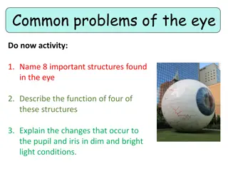
Basic Eye Exam and Visual Acuity Assessment - A Comprehensive Guide
Learn about the 5-point eye exam covering visual acuity, gross examination, motility, intraocular pressure, and funduscopy. Understand how to assess visual acuity at different distances, use the Snellen chart, and manage patients with poor vision. Explore techniques for evaluating near vision in patients aged 35 and older using the Jaeger notation.

Download Presentation

Please find below an Image/Link to download the presentation.
The content on the website is provided AS IS for your information and personal use only. It may not be sold, licensed, or shared on other websites without obtaining consent from the author.If you encounter any issues during the download, it is possible that the publisher has removed the file from their server.
You are allowed to download the files provided on this website for personal or commercial use, subject to the condition that they are used lawfully. All files are the property of their respective owners.
The content on the website is provided AS IS for your information and personal use only. It may not be sold, licensed, or shared on other websites without obtaining consent from the author.
E N D
Presentation Transcript
BASIC EYE EXAM FELICE KATRINA T. RANCHE CLINICAL ASSOCIATE PROFESSOR
THE 5-POINT EYE EXAM Visual Acuity Gross Examination Motility Intraocular Pressure Funduscopy
VISUAL ACUITY (DISTANCE/FAR) Distance visual acuity is taken one eye at a time. Ask the patient to cover the fellow eye with an occluder or his palm. The visual acuity is taken in this sequence: Uncorrected or without spectacles (sc) Corrected or with spectacles (cc) With pinhole (ph), which is the best acuity a patient can have.
USING THE SNELLEN CHART Ask the patient to stand 6m or 20ft from the Snellen chart. Ask him to read the smallest line that he can see. Record this using the Snellen notation. You may use the metric system or the imperial system, as long as you are consistent.
PATIENTS WITH POOR VISION The largest letter is 20/200 or 6/60. If the patient cannot read it, move him forward by 1 meter. VA at that point is 5/60. Move another meter closer if the patient still cannot see. 4/60, 3/60, 2/60, 1/60
PATIENTS WITH POOR VISION If your patient cannot see the E from 1m or 3ft, check if he can COUNT FINGERS (CF or FC) at 3feet. CF at 2ft, 1ft, 6
PATIENTS WITH POOR VISION If your patient cannot count fingers at 6 , check if he can detect HAND MOTIONS (HM) or not. HM is always paired with LIGHT PROJECTION (LPj), which you can test with a penlight. Good = 4 quadrants Fair = 2-3 quadrants Poor = 1 quadrant
PATIENTS WITH POOR VISION If your patient cannot detect hand motions or localize light, check if he can detect the presence of light. Report this as LIGHT PERCEPTION (LP) or NO LIGHT PERCEPTION (NLP).
NEAR VISION Test this in patients aged 35 and older, in whom we expect a decrease in accommodation. Hold the near vision chart 14 inches away and ask the patient to read until the smallest line. Record using the Jaeger notation. Near vision is a binocular function.
SAMPLE REPORTING sc cc ph Near OD 20/100 20/40 20/20 J3 OS 20/70 20/40 20/40
GROSS EXAMINATION Eyelids and eyelashes Proptosis or enophthalmos Corneal clarity and corneal light reflex Sclera Bulbar and palpebral conjunctiva Pupil size and pupillary light reaction
MOTILITY (EOM TESTING) Ask the patient to follow your finger in the 6 directions of gaze. Version - Conjugate binocular eye movement Duction - Monocular eye movement
PALPATION TONOMETRY Ask the patient to look down. Place you index fingers on the right upper eyelid. Press lightly using alternating fingers to check the consistency of the right globe. Repeat for the left. hypotonic: lips soft: ala of nose firm: forehead Do not palpate if you suspect an open eye injury.
DIRECT FUNDUSCOPY Use your ophthalmoscope properly Adjust the rheostat to the brightest light your patient can tolerate Adjust for EOR (red for myopes, green for hyperopes, 0 for plano) Adjust aperture size (smaller than pupil) to reduce glare Ask the patient to look at a distant target Use your right eye to examine the patient s right eye and vice versa
FUNDUSCOPY REPORTING Clear or hazy media Distinct or indistinct disc borders Cup:disc ratio AV ratio Hemorrhages or exudates Good or dull foveal reflex


")


")