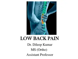
Understanding Low Back Pain Evaluation and Management
This informative resource delves into the evaluation, management, and prognosis of low back pain. It covers evidence-based evaluation, differential diagnosis, and the importance of clinical red and yellow flags in assessing low back pain. Guidelines from the American College of Physicians and the American Pain Society are highlighted, emphasizing the importance of conducting a focused history and physical examination to categorize patients with low back pain. The content also discusses the significance of assessing psychosocial risk factors and provides insights into conducting a thorough neurological examination and classifying patients based on specific criteria.

Download Presentation

Please find below an Image/Link to download the presentation.
The content on the website is provided AS IS for your information and personal use only. It may not be sold, licensed, or shared on other websites without obtaining consent from the author. Download presentation by click this link. If you encounter any issues during the download, it is possible that the publisher has removed the file from their server.
E N D
Presentation Transcript
Welcome to Low Back Pain: Evaluation, Management, and Prognosis
Welcome and Overview Bill McCarberg Founder Chronic Pain Management Program Kaiser Permanente San Diego, California Adjunct Assistant Clinical Professor University of California School of Medicine San Diego, California
Evidence-Based Evaluation of Patients With Low Back Pain
Learning Objective Discuss the differential diagnosis for low back pain (LBP) and the importance of clinical red and yellow flags in evaluation of LBP
Low Back Pain Guidelines In 2007, the American College of Physicians (ACP) and American Pain Society (APS) issued comprehensive joint clinical practice guidelines for diagnosis and treatment of LBP Chou R, et al. Ann Intern Med. 2007;147(7):478-491.
Guideline #1 Clinicians should conduct a focused history and physical examination to help place patients with LBP into 1 of 3 broad categories Nonspecific LBP Back pain potentially associated with radiculopathy or spinal stenosis Back pain potentially associated with another specific spinal cause The history should include assessment of psychosocial risk factors, which predict risk for chronic disabling back pain Strong recommendation Moderate-quality evidence Chou R, et al. Ann Intern Med. 2007;147(7):478-491.
Focused History and Physical Examination Determine presence and level of neurological involvement1,2 Classify patients into 3 broad categories Nonspecific LBP potentially associated with radiculopathy Spinal stenosis Back pain potentially associated with another specific spinal cause Patients with serious or progressive neurologic deficits or underlying conditions requiring prompt evaluation Tumor Infection Cauda equina syndrome Patients with other conditions that may respond to specific treatments Ankylosing spondylitis Vertebral compression fracture 1. Deyo RA, et al. JAMA. 1992;268(6):760-765. 2. Bigos SJ, et al. Acute Low Back Problems in Adults. Clinical Practice Guideline, No. 14; 1994.
Evaluation of Back Pain Site Duration Length of illness Time of onset Spread Mode of onset Quality Precipitating factors Intensity Aggravating factors Frequency Relieving factors Associated features McGuirk BE, et al. In: Ballantyne J, Fishman S and Bonica JJ, eds. Bonica's Management of Pain. 2010:1094-1105.
Epidemiology of Low Back Pain 90% of American adults experience an episode of back pain during their lifetime Of patients who have acute back pain 90% to 95% have a non life-threatening condition Although up to 85% cannot be given an exact diagnosis, nearly all recover within 4 to 6 weeks For 5% to 10% of patients, acute back pain is a manifestation of more serious pathology Vascular catastrophes, malignancy, spinal cord compressive syndromes, and infectious disease processes Winters ME, et al. Med Clin North Am. 2006;90(3):505-523.
What Is Seen in Primary Care Practice? In minority of patients presenting for initial evaluation in primary care setting, LBP is caused by1 Cancer (approximately 0.7% of cases) Compression fracture (4%) Spinal infection (0.01%) Estimates for prevalence of ankylosing spondylitis in primary care patients range from 0.3%1 to 5%2 Spinal stenosis and symptomatic herniated disc are present in about 3% and 4% of patients, respectively Cauda equina syndrome most commonly associated with massive midline disc herniation, but rare Estimated prevalence of 0.04%3 1. Jarvik JG, et al. Ann Intern Med. 2002;137(7):586-597. 2. Underwood MR, et al. Br J Rheumatol. 1995;34(11):1074-1077. 3. Deyo RA, et al. JAMA. 1992;268(6):760-765.
Cost of Low Back Pain LBP is one of top 10 reasons patients seek care from family physicians1 Prevalence of LBP has varied from 7.6% to 37% Peak prevalence between 45 and 60 years of age2 Also reported by adolescents and by adults of all ages 80% of adults seek care at some time for acute LBP3 One-third of US disability costs are due to low back disorders3 Direct costs of diagnosing and treating LBP in United States estimated in 1991 to be $25* billion annually4 Indirect costs, including lost earnings, are even higher4 Proper diagnosis and appropriate treatment of LBP saves healthcare resources, relieves suffering *40 billon in 2008 using Consumer Price Index to compute the relative value of money. 1. AAFP. Facts About Family Practice; 1996. 2. Borenstein DG. Curr Opin Rheumatol. 1997;9(2):144-150. 3. Kuritzky L, et al. Prim Care Rep 1995;1:29-38. 4. Frymoyer JW, et al. Orthop Clin North Am. 1991;22(2):263-271.
Etiology of Low Back Pain Nonspecific LBP Back pain potentially associated with radiculopathy or spinal stenosis Back pain potentially associated with another specific spinal cause Chou R, et al. Ann Intern Med. 2007;147(7):478-491.
Structural Sources of Low Back Pain Muscles of the back1,2 Interspinous ligaments2-4 Zygapophyseal joints5-7 Sacroiliac joint(s)8 Intervertebral discs9-12 Mechanical12 or chemical irritation of dura mater13 1. Kellgren JH. Clin Sci. 1938;3:175-190. 2. Bogduk N. Med J Aust. 1980;2(10):537-541. 3. Kellgren JH. Clin Sci. 1939;4:35-46. 4. Feinstein B, et al. J Bone Joint Surg Am. 1954;36-A(5):981-997. 5. Mooney V, et al. Clin Orthop Relat Res. 1976(115):149-156. 6. McCall IW, et al. Spine (Phila Pa 1976). 1979;4(5):441-446. 7. Fukui S, et al. Clin J Pain. 1997;13(4):303-307. 8. Fortin JD, et al. Spine (Phila Pa 1976). 1994;19(13):1475-1482. 9. Wilberg G. Acta Orthop Scand. 1947;19:211-221. 10. Falconer MA, et al. J Neurol Neurosurg Psychiatry. 1948;11(1):13-26. 11. Kuslich SD, et al. Orthop Clin North Am. 1991;22(2):181-187. 12. O'Neill CW, et al. Spine (Phila Pa 1976). 2002;27(24):2776-2781. 13. El-Mahdi MA, et al. Neurochirurgia (Stuttg). 1981;24(4):137-141.
Causes of Low Back Pain Possible sources of back pain have been demonstrated; causes have been more elusive Refuted: conditions traditionally considered to be possible causes are actually not causes Eg, spondylolysis, spondylolisthesis, degenerative changes (spondylosis) Accepted: tumors and infections Untested: muscle sprain, ligament sprain, segmental dysfunction, and trigger points Known source, unknown cause: sacroiliac joints, zygapophyseal joints, internal disc disruption McGuirk BE, et al. In: Ballantyne J, Fishman S and Bonica JJ, eds. Bonica's Management of Pain. 2010:1105-1122.
Diagnostic Triage Guides Subsequent Decision-Making Inquire about Location of pain Frequency of symptoms Duration of pain History of previous symptoms, treatment, and response to treatment Consider possibility of LBP due to problems outside the back Pancreatitis Nephrolithiasis Aortic aneurysm Systemic illnesses (eg, endocarditis or viral syndromes)
Differential Diagnosis for Acute Low Back Pain Patient Age (Years) Disease or Condition Location of Pain Low back, buttock, posterior thigh Aggravating or Relieving Factors Quality of Pain Signs Increased with activity or bending Local tenderness, limited spinal motion Back strain 20-40 Ache, spasm Sharp, shooting, or burning pain; paresthesia in leg Decreased with standing; increased with bending or sitting Increased with walking, especially up an incline; decreased with sitting Positive straight leg raise test, weakness, asymmetric reflexes Mild decrease in extension of spine; may have weakness or asymmetric reflexes Exaggeration of the lumbar curve, palpable step off (defect between spinous processes), tight hamstrings Decreased back motion, tenderness over sacroiliac joints Fever, percussive tenderness; may have neurologic abnormalities or decreased motion May have localized tenderness, neurologic signs, or fever Acute disc herniation Low back to lower leg 30-50 Low back to lower leg; often bilateral Ache, shooting pain, pins and needles sensation Osteoarthritis or spinal stenosis 30-50 Back, Increased with activity or bending Spondylolisthesis Any age Ache posterior thigh Sacroiliac joints, lumbar spine Ankylosing spondylitis 15-40 Ache Morning stiffness Lumbar spine, sacrum Infection Any age Sharp pain, ache Varies Dull ache, throbbing pain; slowly progressive Affected bone(s) Increased with recumbency or cough Malignancy >50 Adapted from: Patel AT, et al. Am Fam Physician. 2000;61(6):1779-1786.
Guideline #2 Clinicians should not routinely obtain imaging or other diagnostic tests in patients with nonspecific LBP Strong recommendation Moderate-quality evidence Chou R, et al. Ann Intern Med. 2007;147(7):478-491.
Plain X-Rays for Low Back Pain There is no evidence that routine plain radiography in patients with nonspecific LBP is associated with a greater improvement in patient outcomes than selective imaging1-3 Exposure to unnecessary ionizing radiation should be avoided, particularly in young women (amount of gonadal radiation from obtaining a single plain radiograph [2 views] of the lumbar spine is equivalent to daily chest radiograph for more than 1 year)4 Routine advanced imaging (computed tomography [CT] or magnetic resonance imaging [MRI]) is not associated with improved patient outcomes,5 identifies radiographic abnormalities poorly correlated with symptoms,6 and could lead to additional, possibly unnecessary interventions7,8 1. Deyo RA, et al. Arch Intern Med. 1987;147(1):141-145. 2. Kendrick D, et al. BMJ. 2001;322(7283):400-405. 3. Kerry S, et al. Br J Gen Pract. 2002;52(479):469-474. 4. Jarvik JG. Neuroimaging Clin N Am. 2003;13(2):293-305. 5. Gilbert FJ, et al. Radiology. 2004;231(2):343-351. 6. Jarvik JG, et al. Ann Intern Med. 2002;137(7):586-597. 7. Jarvik JG, et al. JAMA. 2003;289(21):2810-2818. 8. Lurie JD, et al. Spine (Phila Pa 1976). 2003;28(6):616-620.
Plain X-Rays for Low Back Pain (cont.) Plain radiography is recommended for initial evaluation of possible vertebral compression fracture in select high-risk patients, such as those with a history of osteoporosis or steroid use1 Evidence to guide optimal imaging strategies is not available for LBP that persists for more than 1 to 2 months if there are no symptoms suggesting radiculopathy or spinal stenosis, although plain radiography may be a reasonable initial option (see recommendation 4 for imaging recommendations in patients with symptoms suggesting radiculopathy or spinal stenosis)2 Thermography and electrophysiologic testing are not recommended for evaluation of nonspecific LBP 1. Jarvik JG, et al. Ann Intern Med. 2002;137(7):586-597. 2. Chou R, et al. Ann Intern Med. 2007;147(7):478-491.
Guideline #3 Clinicians should perform diagnostic imaging and testing for patients with LBP when severe or progressive neurologic deficits are present or when serious underlying conditions are suspected on the basis of history and physical examination Strong recommendation Moderate-quality evidence Chou R, et al. Ann Intern Med. 2007;147(7):478-491.
CT or MRI Diagnostic Imaging Prompt work-up with MRI or CT is recommended if severe or progressive neurologic deficits or suspected serious underlying condition; delayed diagnosis and treatment associated with poorer outcomes1-3 MRI is generally preferred over CT if available; does not use ionizing radiation, provides better visualization of soft tissue, vertebral marrow, and the spinal canal4 1. Loblaw DA, et al. J Clin Oncol. 2005;23(9):2028-2037. 2. Todd NV. Br J Neurosurg. 2005;19(4):301-306. 3. Tsiodras S, et al. Clin Orthop Relat Res. 2006;444:38-50. 4. Jarvik JG, et al. Ann Intern Med. 2002;137(7):586-597.
CT or MRI Diagnostic Imaging (cont.) There is insufficient evidence to guide diagnostic strategies in patients who have risk factors for cancer but no signs of spinal cord compression Proposed strategies generally recommend plain radiography or measurement of erythrocyte sedimentation rate3, with MRI reserved for patients with abnormalities on initial testing1,2 Alternative strategy is to directly perform MRI in patients with a history of cancer, the strongest predictor of vertebral cancer;2 for patients older than 50 without other risk factors for cancer, delaying imaging while offering standard treatments and reevaluating within 1 month may also be a reasonable option4 1. Jarvik JG, et al. Ann Intern Med. 2002;137(7):586-597. 2. Joines JD, et al. J Gen Intern Med. 2001;16(1):14-23. 3. van den Hoogen HM, et al. Spine (Phila Pa 1976). 1995;20(3):318-327. 4. Suarez-Almazor ME, et al. JAMA. 1997;277(22):1782-1786.
Guideline #4 Clinicians should evaluate patients with persistent LBP and signs or symptoms or radiculopathy or spinal stenosis with MRI (preferred) or CT only if they are potential candidates for surgery or epidural steroid injection (for suspected radiculopathy) Strong recommendation Moderate-quality evidence Chou R, et al. Ann Intern Med. 2007;147(7):478-491.
Imaging for Low Back Pain The natural history of lumbar disc herniation with radiculopathy in most patients is for improvement within the first 4 weeks with noninvasive management1,2 There is no compelling evidence that routine imaging effects treatment decisions or improves outcomes3 For prolapsed lumbar disc with persistent radicular symptoms despite noninvasive therapy, discectomy or epidural steroids are potential treatment options4-8 Surgery is also a treatment option for persistent symptoms associated with spinal stenosis9-12 1. Vroomen PC, et al. Br J Gen Pract. 2002;52(475):119-123. 2. Weber H. Spine (Phila Pa 1976). 1983;8(2):131-140. 3. Modic MT, et al. Radiology. 2005;237(2):597-604. 4. Gibson JN, et al. Cochrane Database Syst Rev. 2000(3):CD001350. 5. Gibson JN, et al. Cochrane Database Syst Rev. 2005(4):CD001352. 6. Nelemans PJ, et al. Spine (Phila Pa 1976). 2001;26(5):501-515. 7. Peul WC, et al. N Engl J Med. 2007;356(22):2245-2256. 8. Weinstein JN, et al. JAMA. 2006;296(20):2451-2459. 9. Amundsen T, et al. Spine (Phila Pa 1976). 2000;25(11):1424-1435. 10. Atlas SJ, et al. Spine (Phila Pa 1976). 2005;30(8):936-943. 11. Weinstein JN, et al. N Engl J Med. 2007;356(22):2257-2270. 12. Malmivaara A, et al. Spine (Phila Pa 1976). 2007;32(1):1-8.
MRI for Low Back Pain MRI (preferred if available) or CT is recommended for evaluating patients with persistent back and leg pain who are potential candidates for invasive interventions Plain radiography cannot visualize discs or accurately evaluate the degree of spinal stenosis1 However, clinicians should be aware that findings on MRI or CT (such as bulging disc without nerve root impingement) are often nonspecific Recommendations for specific invasive interventions, interpretation of radiographic findings, and additional work-up beyond scope of guideline, but decisions should be based on clinical correlation between symptoms and radiographic findings, severity of symptoms, patient preferences, surgical risks, and costs and will generally require specialist input2 1. Jarvik JG, et al. Ann Intern Med. 2002;137(7):586-597. 2. Chou R, et al. Ann Intern Med. 2007;147(7):478-491.
Critical Clinical Indicators of Pathology In patients with back and leg pain, a typical history for sciatica (back and leg pain in a typical lumbar nerve root distribution) has a fairly high sensitivity, but uncertain specificity for herniated disc1,2 >90% of symptomatic lumbar disc herniations (back and leg pain due to a prolapsed lumbar disc compressing a nerve root) occur at L4/L5 and L5/S1 levels3 1. van den Hoogen HM, et al. Spine (Phila Pa 1976). 1995;20(3):318-327. 2. Vroomen PC, et al. J Neurol. 1999;246(10):899-906. 3. Chou R, et al. Ann Intern Med. 2007;147(7):478-491.
Critical Clinical Indicators of Pathology (cont.) A focused examination that includes straight-leg-raise testing and a neurologic examination that includes evaluation of knee strength and reflexes (L4 nerve root), great toe and foot dorsiflexion strength (L5 nerve root), foot plantarflexion and ankle reflexes (S1 nerve root), and distribution of sensory symptoms should be done to assess the presence and severity of nerve root dysfunction Chou R, et al. Ann Intern Med. 2007;147(7):478-491.
Critical Clinical Indicators of Pathology (cont.) A positive result on straight-leg-raise test (defined as reproduction of the patient s sciatica between 30 and 70 degrees of leg elevation) has a relatively high sensitivity (91% [95% CI, 82% to 94%]), but modest specificity (26% [CI, 16% to 38%]) for diagnosing herniated disc Crossed straight-leg-raise test is more specific (88% [CI, 86% to 90%]), but less sensitive (29% [CI, 24% to 34%]) Deville WL, et al. Spine (Phila Pa 1976). 2000;25(9):1140-1147.
Critical Clinical Indicators of Pathology (cont.) All patients should be evaluated for Presence of rapidly progressive or severe neurologic deficits Motor deficits at more than 1 level, fecal incontinence, and bladder dysfunction Most frequent finding in cauda equina syndrome is urinary retention (90% sensitivity) Without urinary retention, probability is approximately 1 in 10,000 Chou R, et al. Ann Intern Med. 2007;147(7):478-491. Deyo RA, et al. JAMA. 1992;268(6):760-765.
Yellow Flags Identify psychosocial problems in acute phase Slow progress to recovery may be due to undetected, or unrevealed psychosocial factors Pertain to patient's beliefs and behaviors concerning physical activity and domestic, social, and vocational responsibilities Example: patient believes physical activity might harm back, make pain worse, so avoids activities Most destructive is aversion to work Belief that work caused pain, work aggravates pain, work is too heavy, and work should not be done McGuirk BE, et al. In: Ballantyne J, Fishman S and Bonica JJ, eds. Bonica's Management of Pain. 2010:1094-1105.
Psychosocial Factors of Low Back Pain Stronger predictors of LBP outcomes than either physical findings or severity/duration of pain1-3 Assessment of psychosocial factors identifies patients who may have delayed recovery and could help target interventions 1 trial in referral setting found intensive multidisciplinary rehabilitation more effective than usual care in patients with acute or subacute LBP identified as having risk factors for chronic back pain disability4 Direct evidence on effective primary care interventions for identifying and treating such factors in patients with acute LBP is lacking5,6 Evidence is currently insufficient to recommend optimal methods for assessing psychosocial factors and emotional distress7 However, psychosocial factors that may predict poorer LBP outcomes include presence of depression, passive coping strategies, job dissatisfaction, higher disability levels, disputed compensation claims, or somatization8-10 1. Pengel LH, et al. BMJ. 2003;327(7410):323. 2. Fayad F, et al. Ann Readapt Med Phys. 2004;47(4):179-189. 3. Pincus T, et al. Spine (Phila Pa 1976). 2002;27(5):E109-120. 4. Gatchel RJ, et al. J Occup Rehabil. 2003;13(1):1-9. 5. Hay EM, et al. Lancet. 2005;365(9476):2024-2030. 6. Jellema P, et al. BMJ. 2005;331(7508):84. 7. Chou R, et al. Ann Intern Med. 2007;147(7):478-491. 8. Steenstra IA, et al. Occup Environ Med. 2005;62(12):851-860. 9. Deyo RA, et al. Spine (Phila Pa 1976). 2006;31(23):2724-2727. 10. Carey TS, et al. Spine (Phila Pa 1976). 1996;21(3):339-344.
Red Flags of Lower Back Pain History Gradual onset of back pain Age <20 years or >50 years Thoracic back pain Pain lasting longer than 6 weeks History of trauma Fever/chills/night sweats Unintentional weight loss Pain worse with recumbency Pain worse at night Unrelenting pain despite supratherapeutic doses of analgesics History of malignancy History of immunosuppression Recent procedure causing bacteremia History of intravenous drug use Physical Examination Fever Hypotension Extreme hypertension Pale, ashen appearance Pulsatile abdominal mass Pulse amplitude differentials Spinous process tenderness Focal neurologic signs Acute urinary retention Winters ME, et al. Med Clin North Am. 2006;90(3):505-523.
Risk for Chronicity Vertebral infection Intravenous drug use, recent infection Vertebral compression fracture Older age, history of osteoporosis, and steroid use Musculoskeletal Inactivity In general Emotional distress
Cancer-Related Risk Factors Large, prospective study from a primary care setting History of cancer (positive likelihood ratio, 14.7) Unexplained weight loss (positive likelihood ratio, 2.7) Failure to improve after 1 month (positive likelihood ratio, 3.0) Age >50 years (positive likelihood ratio, 2.7) Posttest probability of cancer increases from approximately 0.7% to 9% in patients with a history of cancer (not including nonmelanoma skin cancer) In patients with any 1 of the other 3 risk factors, the likelihood of cancer only increases to approximately 1.2% Deyo RA, et al. J Gen Intern Med. 1988;3(3):230-238.
Non-Cancer-Related Risk Factors Features predicting vertebral infection not well studied, but may include fever, intravenous drug use, or recent infection1 Consider risk factors for vertebral compression fracture, such as older age, history of osteoporosis, and steroid use; and for ankylosing spondylitis, such as younger age, morning stiffness, improvement with exercise, alternating buttock pain, and awakening due to back pain during the second part of the night only2 Clinicians should be aware that criteria for diagnosing early ankylosing spondylitis (before the development of radiographic abnormalities) are evolving3 1. Jarvik JG, et al. Ann Intern Med. 2002;137(7):586-597. 2. Rudwaleit M, et al. Arthritis Rheum. 2006;54(2):569-578. 3. Rudwaleit M, et al. Arthritis Rheum. 2005;52(4):1000-1008.
Racial/Cultural Aspects of Assessment To communicate effectively with all patients Always use simple words, not medical jargon Determine what the patient/caregiver already knows or believes about his/her health situation Encourage questions by asking, What questions do you have? (allows for an open- ended response), instead of Do you have any questions? (allows for a no response, ending the conversation) Use the teach-back method to confirm the level of understanding: Ask patients/family members to restate what was just communicated in the appointment or meeting Zacharoff KL. Cross-Cultural Pain Management: Effective Treatment of Pain in the Hispanic Population; 2009.
Culturally Competent Care Ensure that patients/consumers receive effective, understandable, and respectful care that is provided in a manner compatible with their cultural health beliefs and practices and preferred language Implement strategies to recruit, retain, and promote at all levels of the organization a diverse staff and leadership that are representative of the demographic characteristics of the service area Ensure that staff, at all levels and across all disciplines, receives ongoing education and training in CLAS delivery USDHHS OMH. National Standards for Culturally and Linguistically Appropriate Services (CLAS) in Health Care; 2001.
Avoiding Racial and Cultural Bias per Knox H. Todd, MD, MPH Make pain assessment mandatory Give a nonopioid analgesic at triage Track reasons for unscheduled returns Audit for ethnic bias Consider which pain scales should be used Use multilingual laminated cards Todd KH. Medical Ethics Advisor. 1999.
Pearls for Practice Categorize patients into 1 of 3 broad groups: nonspecific low back pain, back pain potentially associated with radiculopathy or spinal stenosis, or back pain potentially associated with another specific spinal cause Evaluate psychosocial risk factors to predict the risk for chronic, disabling low back pain Provide patients with evidence-based information on expected course of low back pain, effective self-care options, and recommend that they be physically active Chou R, et al. Ann Intern Med. 2007;147(7):478-491.
Questions? Please pass your question card to a staff member.
Treatment of Low Back Pain: Pharmacologic and Nonpharmacologic Options Roger Chou, MD, FACP Associate Professor of Medicine, Department of Medicine Department of Medical Informatics and Clinical Epidemiology Oregon Health & Science University
Disclosure: Roger Chou, MD, FACP Dr. Chou has disclosed that he has no actual or potential conflict of interest in regard to this activity His presentation will include off-label discussion of anticonvulsants, benzodiazepines, and tricyclic antidepressants for the treatment of low back pain (LBP)
Learning Objective Integrate evidence-based pharmacologic and nonpharmacologic therapies into a comprehensive treatment plan for chronic LBP
Low Back Pain Burden LBP is the fifth most common reason for US office visits, and the second most common symptomatic reason1-2 $90.7 billion dollars in total healthcare expenditures in 19983 LBP is the most common cause for activity limitations in persons under the age of 454 1. Hart LG, et al. Spine (Phila Pa 1976). 1995;20(1):11-19. 2. Deyo RA, et al. Spine (Phila Pa 1976). 2006;31(23):2724-2727. 3. Luo X, et al. Spine (Phila Pa 1976). 2004;29(1):79-86. 4. Von Korff M, et al. Spine (Phila Pa 1976). 1996;21(24):2833-2837.
Increasing Rates of Back Surgery Trends in Rates of Discectomy/Laminectomy and Fusion in 1992-2003 US Average Rate of Discharges per 1000 Medicare Enrollees Weinstein JN, et al. Spine (Phila Pa 1976). 2006;31(23):2707-2714.
Increasing Rates of Back Injections Lumbosacral Injection Rates by Year: Age- and Sex-Adjusted per 100,000 2055.2 553.4 263.9 212.3 79.7 SI=sacroiliac. Friedly J, et al. Spine (Phila Pa 1976). 2007;32(16):1754-1760.
Increasing Costs Mean ($) Year Martin BI, et al. JAMA. 2008;299(6):656-664.
Rising Prevalence of Chronic LBP Prevalence of Chronic Low Back Pain in North Carolina, 1992 and 2006 % Prevalence (95% CI) 1992: 3.9% 2006: 10.2% 1992 (n=8067) 3.9 (3.4-4.4) 2006 (n=9924) 10.2 (9.3-11.0) PRR Characteristic Total Sex Male Female Age (Years) 21-34 35-44 45-54 55-64 65 Race/Ethnicity Non-Hispanic White Non-Hispanic Black Hispanic Other % Increase 162 (2.5-97.5% CI)* 2.62 (2.21-3.13) 2.9 (2.2-3.6) 4.8 (4.0-5.6) 8.0 (6.8-9.2) 12.2 (10.9-13.5) 176 154 2.76 (2.11-3.75) 2.54 (2.13-3.08) 1.4 (0.8-2.0) 4.8 (3.3-6.3) 4.2 (3.0-5.5) 6.3 (4.2-8.3) 5.9 (4.5-7.3) 4.3 (3.0-5.6) 9.2 (7.2-11.2) 13.5 (11.4-15.7) 15.4 (12.8-17.9) 12.3 (10.2-14.4) 201 92 219 146 109 3.01 (1.95-5.17) 1.92 (1.35-2.86) 3.19 (2.29-4.59) 2.46 (1.73-3.50) 2.09 (1.62-2.84) 4.1 (3.5-4.7) 3.0 (2.0-4.0) ** 4.1 (1.4-6.8) 10.5 (9.4-11.5) 9.8 (8.2-11.4) 6.3 (3.8-8.9) 9.1 (6.0-12.0) 155 226 2.55 (2.13-3.05) 3.26 (2.32-4.96) 120 2.20 (1.16-6.99) CI=confidence interval. PRR=prevalence rate ratio. *The PRRs and CI were estimated via bootstrapping; 97.5% CIs were reported rather than to assume normality. **Unable to estimate owing to scall cell count (n<5). Freburger JK, et al. Arch Intern Med. 2009;169(3):251-258.
Practice Patterns Spine surgery rates in the US are the highest in the world Rates in the US 5 times higher than in the UK 20-fold variation in fusion: 4.6 per 1000 in Idaho Falls to 0.2 per 1000 in Bangor, Maine Interventional therapies are also widely used Intradiscal electrothermal therapy estimated at 7000-10,000 annually 20-fold variation in epidural steroid injections: 104 per 1000 in Palm Springs to 5.6 per 1000 in Honolulu Deyo RA, et al. Clin Orthop Relat Res. 2006;443:139-146. Weinstein JN, et al. Spine (Phila Pa 1976). 2006;31(23):2707-2714.
7 Back Pain Breakthroughs: Are you hurting? Here s help. Reader s Digest July 2007 End Back Pain Agony (Michael J. Weiss) Weiss MJ. Reader's Digest. July, 2007.


")


")