Understanding the Human Eye: Structure and Function

The human eye is a remarkable optical system that functions like a camera, with components such as the cornea, lens, and retina working together to form images that are perceived and interpreted by the brain. The structure of the eye, including the cornea, iris, lens, and retina, plays a crucial role in capturing and processing light to create visual perceptions. By understanding the intricate workings of the eye, we can appreciate its complexity and importance in our daily lives.

Uploaded on Sep 26, 2024 | 0 Views
Download Presentation

Please find below an Image/Link to download the presentation.
The content on the website is provided AS IS for your information and personal use only. It may not be sold, licensed, or shared on other websites without obtaining consent from the author. Download presentation by click this link. If you encounter any issues during the download, it is possible that the publisher has removed the file from their server.
E N D
Presentation Transcript
EYE AND EAR VISHAL KUMAR ASSISTANT PROFESSOR CSJMU
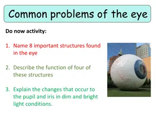
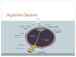
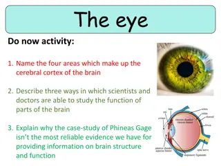
Eye structure The human eye is a complex optic instrument. Its main goal is to transfer the correct image to the optic nerve. Cornea is a transparent coat covering the front part of the eye. It has no blood vessels. Cornea borders sclera which is a non-transparent eye coat. Anterior chamber is a space between cornea and iris. It is filled with intra-ocular fluid. Iris looks like a circle with an opening in the middle (pupil). Iris consist of muscles that change pupil size by constricting and relaxing. Iris is responsible for the colour of the eyes (if it is blue this means it contains few pigment cell, if brown a lot). Its function is same as of aperture in a camera to adjust light flow. Pupil is an aperture in iris. Its size usually depends on the illumination level. The more light the smaller the pupil. Crystalline lens is the eye natural lens'. It is transparent, elastic can change its shape, focusing in almost instantly, therefore one can see well both near and far. It is located in a capsule and is with held by ciliary muscle. Vitreous body is a gel-like transparent substance located in the posterior part of the eye. The vitreous body supports the sphere of the eye ball. Retina consists of photoreceptors and nerve cells. There are two types of receptor cells in retina: cones and rods. These cells producing rhodopsin enzyme transform light energy into electric energy of neural tissue. Rods have high light sensitivity and allow seeing in poor light, they are also responsible for periphery vision. Cones need plenty of light for functioning but allow to distinguish small details and ensure colour appreciation. Most cones are located in macula which is responsible for the sharpest vision. Sclera is the non-transparent outer coat of the eye and in the frontal part of the eye it verges into the transparent cornea. 6 eye moving muscles are attached to it. It contains a few nerve terminals and vessels. Choroidinlays the back part of sclera, it adjoins retina and is closely linked to it. Choroid is responsible for blood supply of intraocular structures. Choroid has no nerve terminals therefore when there is a trouble there, there is no pain which usually alarms about a problem. Optic nerve transfers signals from nerve terminals to the brain.
Function of the Human Eye The human eye is a complex optical system that basically works like a camera: the iris serves as the aperture that controls the amount of light rays reaching cornea and lens (photographic objective), and the retina works as the film. Bending of light rays by cornea and lens serves to create sharp images on the retina. These images ultimately trigger nerve impulses, which are transmitted to the brain where the images are perceived and interpreted. The human eye also focuses and lets in light to produce images.So basically,light rays that are deflected from or by distant objects land on the retina after they pass through various mediums like the cornea, crystalline lens, aqueous humor,the lens,and vitreous humor. The concept here though is that as the light rays move through the various mediums, they experience refraction of light. Well,to put it in simple terms,refraction is nothing but the change in direction of the rays of light as they pass between different mediums. Having different refractive indexes is what bends the rays to form an image. The light rays finally are received and focused on the retina. The retina contains photoreceptor cells called rods and cones and these basically detect the intensity and the frequency of the light. Further, the image that is formed is processed by millions of these cells, and they also relay the signal or nerve impulses to the brain via the optic nerve. The image formed is usually inverted but the brain corrects this phenomenon. This process is also similar to that of a convex lens.
STRUCTURE OF EAR What are the parts of the ear? The three main parts of your ear include the outer ear, middle ear and inner ear. Your tympanic membrane (eardrum) separates your outer ear and middle ear. Outer ear (external ear) Your outer ear is the part of your ear that s visible. It s what most people mean when they say ear. Also called the auricle or pinna, your outer ear consists of ridged cartilage and skin, and it contains glands that secrete ear wax. Its funnel-shaped canal leads to your eardrum, or tympanic membrane. Middle ear Your middle ear begins on the other side of your tympanic membrane (eardrum). There are three tiny bones in this area the malleus, incus and stapes. They transfer sound vibrations from your eardrum to your inner ear. Your middle ears also house the eustachian tubes, which help equalize the air pressure in your ears. Inner ear Your inner ear contains two main parts: the cochlea and the semicircular canals. Your cochlea is the hearing organ. This snail-shaped structure contains two fluid-filled chambers lined with tiny hairs. When sound enters, the fluid inside of your cochlea causes the tiny hairs to vibrate, sending electrical impulses to your brain. . The snail- like cochlea is made up of three fluid-filled chambers that spiral around a bony core, which contains a central channel called the cochlear duct. Inside the cochlear duct is the main hearing organ, the spiral shaped organ of Corti. Hair cells inside the organ of Corti detect sound and send the information through the cochlear nerve. Sound waves enter through the outer ear, move into the middle ear, and finally reach the inner ear and its intricate network of nerves, bones, canals, and cells. The semicircular canals, also known as the labyrinthine, are responsible for balance. They tell your brain which direction your head is moving.
FUNCTION OF EAR What is the main function of the ear? Your ears have two main functions: hearing and balance. Hearing: When sound waves enter your ear canal, your tympanic membrane (eardrum) vibrates. This vibration passes on to three tiny bones (ossicles) in your middle ear. The ossicles amplify and transmit these sound waves to your inner ear. Once the sound waves reach your inner ear, tiny hair cells called stereocilia transform the vibrations into electrical energy and send it along nerve fibers to your brain. Balance: Your inner ear contains semicircular canals filled with fluid and hair-like sensors. When you move your head, the fluid inside these loop-shaped canals sloshes around and moves the hairs. The hairs transmit this information along the vestibular nerve to your brain. Finally, your brain sends signals to your muscles to help you stay balanced.