Understanding Mosaicism: Types, Causes, and Implications in Genetics
Mosaicism is the presence of genetically different cell lines within one individual, often involving sex chromosomes due to mitotic defects in early development. Chromosomal mosaicism can be distinguished from chimerism, and its association with gametogenesis and aneuploidy is influenced by maternal age. Errors in meiosis play a significant role in the occurrence of mosaicism, emphasizing the importance of understanding its mechanisms and implications in genetic disorders.

Download Presentation

Please find below an Image/Link to download the presentation.
The content on the website is provided AS IS for your information and personal use only. It may not be sold, licensed, or shared on other websites without obtaining consent from the author. Download presentation by click this link. If you encounter any issues during the download, it is possible that the publisher has removed the file from their server.
E N D
Presentation Transcript
Mosaicism Lecturer PhD. Asmaa Amer Almukhtar Medical Genetics Department/ICCMGR Nov 2023
Normal karyotype What is the mosaicism? Who is the responsible about it? Types of mosaicism. How it creates?
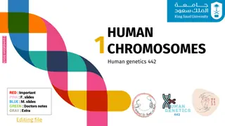
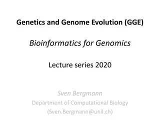
Normal female human karyotype 46,XX Normal Male Human Karyotype 46,XY
Mosaicism is the possession of multiple genetically different cell lines multiple genetically different cell lines in a single person in a single person. Most chromosomal mosaicism involves the sex chromosomes and occurs because of defects in mitosis in an early embryo. Normally, chromosomes duplicate and separate equally in mitotic division. Mosaicism can occur when the chromosomes fail to separate (mitotic nondisjunction) or fail to migrate (anaphase lag). In general, the greater the proportion of abnormal cell lines the greater the proportion of abnormal cell lines, the more abnormal the phenotype abnormal the phenotype. The earlier in embryonic earlier in embryonic development an abnormal cell is established, the higher the percentage of abnormal cells the higher the percentage of abnormal cells in that person. in that person.
Chromosomal mosaicism Chromosomal mosaicismis the presence of two (or more) cell lines withdifferent differentkaryotypes, originatingfrom a single zygote. must be distinguished from the so-called chimera, lines with different karyotypes different karyotypes, but they come from two zygotes humans, we know the the 46 46,XX/ ,XX/46 46,XY chimera, ,XY chimera,a very rare genetic associated withtrue hermaphroditism, true hermaphroditism,probably most chimeras escape detection from a single zygote.The mosaic chimera,which contains two cell from two zygotes. In
The association between gametogenesis and mitosis after fertilization It is well known that uniform aneuploidy, which is derived from meiotic chromosomal malsegregation, is critically dependent on maternal age maternal age. The current mainstream thinking believes that errors in maternal meiosis errors in maternal meiosis, especially the MI, are the main factors leading to aneuploidy ,while the paternal meiosis errors only account for the paternal meiosis errors only account for 1 1% % .The reason can be attributed to the following: there is a rigorous checkpoint in the process of male there is a rigorous checkpoint in the process of male spermatogenesis spermatogenesis, which can effectively avoid the development of abnormal chromosomes while in the process of while in the process of oogenesis, such checkpoints are often lacking or not strict enough. oogenesis, such checkpoints are often lacking or not strict enough. Further, in the process of oogenesis, oocytes are stored in follicular pools since the fetal period and arrested in the prophase of meiosis I until ovulation occurs years later. During this long During this long- -term period, cohesion of sister chromatids is deteriorated with maternal age term period, cohesion of sister chromatids is deteriorated with maternal age ..Thus, oogenesis is more prone to errors than spermatogenesis.
autosometrisomies(trisomy 21, 13, 18) occur less often in mosaicis.trisomy trisomy 8 8is also known , which occursonly in mosaic form live births . Mosaic always arisespostzygotic, during mitotic division or loss of a chromosome during division of a trisomic or normal zygote. The ratio of cell lineages then depends on which division the nondisjunction or loss of the chromosome occurred in and on how viable the abnormal cells are. E.g. postzygotic nondisjunction or loss of a chromosome affecting the autosomes would lead to the extinction of the monosomic line (monosomy of the autosomes is lethal even at the cellular level lethal even at the cellular level). Mosaicism resulting from the loss of one chromosome from a the loss of one chromosome from a trisomic of normal and of normal and trisomic trisomiccell lines. cell lines. only in mosaic formin postzygotic,i.e. nondisjunction Mosaicism resulting from trisomic zygote results in a zygote results in amosaic mosaic
Non Non- -disjunction disjunction Non Non- -disjunction is the failure of sister chromatids to separate during mitosis disjunction is the failure of sister chromatids to separate during mitosis. Instead of separating, the entire chromosome (two chromatids) is pulled to one cell (two chromatids) is pulled to one cell, creating a cell with a monosomy and another cell with a trisomy . The degree of mosaicism(the percentage) depend on the time of non disjunction The degree of mosaicism(the percentage) depend on the time of non disjunction Instead of separating, the entire chromosome Anaphase lagging Anaphase lagging Anaphase lagging is the failure of a single chromatid to be incorporated into the nucleus resulting in a monosomy of that Anaphase lagging is the failure of a single chromatid to be incorporated into the nucleus resulting in a monosomy of that chromosome in one cell and a disomy in the corresponding chromosome in the other cel chromosome in one cell and a disomy in the corresponding chromosome in the other cell. Anaphase lagging occurs when the chromatid fails to attach to the spindle or when the chromatid attaches to the spindle but then fails to be incorporated in the nucleus. If this mechanism occurs prior to differentiation, then the organism will contain two distinct cell lines, thereby creating a general mosaic. If this event occurs after differentiation in the trophoblast, then the placenta will contain a normal and monosomic cell line, an example of CPM. Endoreplication Endoreplication Endoreplication is the replication of a chromosome without division Endoreplication is the replication of a chromosome without division. This results in a trisomic disomic chromosome in the other. disomic chromosome in the other. Chromosome gain is believed to derive from two mechanisms, a cell cycle malfunction in which a chromosome is replicated without subsequent cytokinesis or when mitosis is initiated and shortly thereafter shutdown, resulting in a replicated chromosome. Regardless of the mechanism, the result is the same: the gain of a single chromosome (Fig.2E). Another name for endoreplication is polyploidy endoreplication is polyploidy. Polyploidy has been shown to exist in blood, gut, skin and brain . trisomic chromosome in one cell and a chromosome in one cell and a
Uniparental disomy Uniparental disomy Non-disjunction, anaphase lagging and endoreplication lead to aneuploidy. However, there are mitotic events that lead to mosaicism, but still present a disomic cell line. Instead of chromosomes presenting in a gain or loss fashion, a phenomenon known as uniparental disomy (UPD) can occur. As UPD implies, there are two chromosomes present; however, instead of one maternal and paternal chromosome, there are two copies of either a chromosomes present; however, instead of one maternal and paternal chromosome, there are two copies of either a maternal or paternal chromosomes. This may be the result of a maternal or paternal chromosomes. This may be the result of a trisomic (Fig.2F). The most frequent chromosome that UPD occurs in is chromosome 15, where two paternal copies is referred to as Angelman syndrome or two maternal copies is known as Prader-Willi syndrome. Other chromosomes that can present with UPD are chromosomes 7, 11 and 16 (Kaluoseket al., 1992). As UPD implies, there are two trisomic rescue event after an error during meiosis rescue event after an error during meiosis Abnormalities in spindles are also more prevalent in older women. Abnormalities in spindles are also more prevalent in older women. The centrosome is inherited from the sperm and is responsible for the first mitotic divisions within the human embryo The disruption of the sperm centrosome can produce The disruption of the sperm centrosome can produce mosaicism in the preimplantation embryo mosaicism in the preimplantation embryo
Different mechanisms leading to chromosome malsegregation in humans. For each figure, two different chromosomes are present, black chromosomes are paternal in origin and white are maternal in origin. (A A) Proper segregation of chromosomes during mitosis. (B B) A mitotic non-disjunction event in a paternal chromosome. (C C) An anaphase lagging event involving a paternal chromosome. (D D) A trisomy rescue event involving a paternal chromosome. (E E) An endoreplication event involving a paternal chromosome. (F F) A trisomy rescue event with uniparental disomy in the paternal chromosomes.
Mosaicism limited to the placenta Mosaicism may be limited to the placenta . The literature reports that a relatively high percentage (1-2%) of first-trimester embryos are mosaics of trisomic and disomic (normal) lines, which was found when examining chorionic villus cells. Because mosaicism is not confirmed in most fetuses, such mosaicism is called confined placental mosaicism confined placental mosaicism ( CPM =confined placental mosaicism). However, some of these fetuses or neonates may show uniparental disomy.
Pseudomosaicism Duringprenatal cytogenetic examination we can encounter so- called pseudomosaicism pseudomosaicism..Unlike true mosaicism, which is actually present in the cells of an individual, pseudomosaicism arises when cells are cultivated in tissue culture. are cultivated in tissue culture. arises when cells
Lyonization Inactivation of the X chromosome Inactivation of the X chromosomeorlyonization the stage of anembryoconsisting of 100 (most often in the case of a normal female karyotype 46,XX; however, it also occurs in male individuals sex withKlinefelter syndrome - karyotype 47,XXY and in other pathological karyotypes with more than one X chromosome so that in the final state there is only one active X chromosome in the cell). Inactivation of the X chromosome is random chromosome is randomin each cell of the embryo , but also permanent, division of this cell will already have the same inactivated chromosome, whether of maternal or paternal origin. The inactivated X chromosome in this way represents a deposit of highly condensed chromatin, as the so-calledBarr body Barr bodyor sex chromatin. sex chromatin.Individuals with monosomy 45,X, as well as 46,XY males, do not have a Barr body. The inactivation process is controlled by a regulatory region known as the X X- -inactivation center (XIC). center (XIC).In this region there is, among other things, the gene for non-coding RNAXIST specific transcript (non-protein coding); Xq13.2;and several of its regulators including the TSIX transcript, XIST antisense RNA; Xq13.2;.It is the RNA product of the XIST gene that induces changes in the conformation of the X chromosome, which ultimately lead to its inactivation. Genesstored in thepseudoautosomal regionof the X chromosome are not inactivated. The inactivation of the X chromosome is also called the Lyonization process the Lyonization processin honor of the British geneticistMary Frances Lyon Mary Frances Lyon(1925-2014), who first described this process in 1961. lyonizationoccurs in the early stages of development (approximately at 100- -200 200 cells cells) if thekaryotypecontains more than one X chromosome Inactivation of the X permanent,because all other cells arising from the chromatin,visible inactivation XIST( X inactive TSIXgene ( TSIX


 occur less often")