Understanding Dry Eye Disease: Causes, Symptoms, and Management
Dry eye disease is a common condition caused by factors leading to inflammation of the eye and tear-producing glands, affecting tear production and eye lubrication. This presentation covers the definition, clinical features, and management of dry eye, emphasizing its impact on quality of life and importance of proper diagnosis and treatment.

Download Presentation

Please find below an Image/Link to download the presentation.
The content on the website is provided AS IS for your information and personal use only. It may not be sold, licensed, or shared on other websites without obtaining consent from the author. Download presentation by click this link. If you encounter any issues during the download, it is possible that the publisher has removed the file from their server.
E N D
Presentation Transcript
Dry Eye Dr.Ajai Agrawal Additional Professor Department of Ophthalmology AIIMS, Rishikesh 1
Acknowledgement Photographs in the presentation are courtesy of Dr.Brad Bowling (Kanski s Clinical Ophthalmology) 2
Learning Objectives At the end of this class the students shall be able to : Define dry eye disease. Understand predisposing and aetiological factors responsible for dry eye disease Comprehend clinical features and tests for the above condition Understand fundamentals of managing dry eye depending on the severity of disease 3
What is Dry Eye Disease? Dry eye disease (DED) is a condition caused by many factors that result in inflammation of the eye and tear-producing glands. Inflammation can decrease the ability of the eye to produce normal tears that protect the surface of the eye and keep it moist and lubricated. 4
Definition Dry eye is not a trivial complaint. It can cause significant discomfort and affect quality of life significantly. In 1995 the National Eye Institute defined dry eye disease (DED) as a disorder of the tear film due to tear deficiency or excessive tear evaporation which causes damage to the interpalpebral ocular surface and is associated with symptoms of ocular discomfort . 5
Definition In 2007 the International Dry Eye Workshop defined it as a multifactorial disease of the tears and ocular surface that results in symptoms of discomfort, visual disturbance, and tear film instability with potential damage to the ocular surface. It is accompanied by increased osmolarity of the tear film and inflammation of the ocular surface. 6
Dry Eye Affects Quality of Life 8
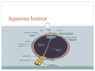
The Healthy Eye Normal tearing depends on a neuronal feedback loop Secretomotor Nerve Impulses Lacrimal Glands Tears Support and Maintain Ocular Surface Ocular Surface Neural Stimulation 9
Dry Eye Disease: An Immune-Mediated Inflammatory Disorder Inflammation disrupts normal neuronal control of tearing Lacrimal Glands: Neurogenic Inflammation T-cell Activation Cytokine Secretion into Tears Interrupted Secretomotor Nerve Impulses Tears Inflame Ocular Surface Cytokines Disrupt Neural Arc 10
Multiple Factors in Dry Eye Transient discomfort May be stimulated by environmental conditions Inflammation and ocular surface damage Altered tear film composition 1de Paiva and Pflugfelder. In: Dry Eye and Ocular Surface Disorders. 2004; 2Pflugfelder et al. In: Dry Eye and Ocular Surface Disorders. 2004. 11
Role of Inflammation in Chronic Dry Eye Inflammation may be present but not clinically apparent Cycle of inflammation and dysfunction If untreated, inflammation can damage lacrimal gland and ocular surface Consequences: Lower tear production Altered corneal barrier function 12 Pflugfelder. Am J Ophthalmol. 2004.
Healthy Tears A complex mixture of proteins, mucin, and electrolytes Antimicrobial proteins: Lysozyme, lactoferrin Growth factors & suppressors of inflammation: EGF, IL-1RA Soluble mucin secreted by goblet cells for viscosity Electrolytes for proper osmolarity Stern et al. In: Dry Eye and Ocular Surface Disorders. 2004. 13 Image adapted from: Dry Eye and Ocular Surface Disorders. 2004.
Tears in Chronic Dry Eye Decrease in many proteins Decreased growth factor concentrations Altered cytokine balance promotes inflammation Soluble mucin 5AC greatly decreased Due to goblet cell loss Impacts viscosity of tear film Proteases activated Increased electrolytes Solomon et al. Invest Ophthalmol Vis Sci. 2001. Zhao et al. Cornea. 2001. Ogasawara et al. Graefes Arch Clin Exp Ophthalmol. 1996. Image adapted from: Dry Eye and Ocular Surface Disorders. 2004. 14
Who Is Likely to Have Dry Eye? How Do We Diagnose It? 15
Dry Eye: Multifactorial nature Elderly woman Taking glaucoma medications Post menopausal Contact lens user Working for long hours in front of computer Air-conditioned environment 16
Patient Types with High Incidence of Dry Eye Disease Women aged 50 or older Women using postmenopausal hormone replacement therapy Those with ocular co-morbidities xerophthalmia, cicatrical pemphigoid, atopic keratoconjunctivitis, ocular rosacea Contact lens wearers Smokers 17
Dry Eye Disease: Predisposing Factors Ageing Menopause - Decreased Androgens Allergy Response Environmental Stresses Contact Lens Wear Wind Air Pollution Ocular Surgery (LASIK, Corneal Transplant) Medications Low Humidity: Heating/AC Lack of Sleep Use of Computer Terminals 18
Medications That May Contribute to Dry Eye Disease Systemic Anti-hypertensives Anti-androgens Anti-cholinergics Antidepressants Cardiac Anti-arrhythmic Drugs Parkinson s Disease Agents Antihistamines Topical Preservatives in Tears 19
Dry Eye Disease: Autoimmune Triggers Systemic Autoimmunity Rheumatoid Arthritis Lupus Sj gren s Syndrome Graft vs. Host Disease All can result in immune-mediated inflammation in the eye. Inflammatory mediators secreted into tears. Promote inflammation of ocular surface. 20
Current Triggers of Dry Eye Disease Environment Medications Contact Lens Surgery Rheumatoid Arthritis Lupus Sj gren s Graft vs Host Inflammation Irritation Tear Deficiency/ Instability Postmenopause Meibomian Gland Disease Symptoms of Ocular Surface Disease 21
Dry Eye Disease Symptoms Discomfort Dryness Burning, Stinging Foreign-Body Sensation Gritty Feeling, Stickiness Blurry Vision Photophobia, Itching, Redness Note: Symptoms seldom correlate with clinical signs 24
Clinical Presentation Can Vary in Severity Mild Severe Slitlamp Fluorescein Dye Stain 25
Slit lamp examination Increased debris/mucin strands in tear film Inspection of tear meniscus at lid margin. Normal thickness 1mm, convex. < 0.5mm tear deficiency. In severe cases Marginal tear meniscus is concave, small & absent. 26
Filaments ( comma shaped) over corneal surface which move on blinking 27
Mucous plaques semi-transparent, white to grey, slightly elevated lesions Stain with rose bengal. 28
Bulbar conjunctival vessels may be dilated Red Eye Corneal surface irregularity/ dry areas. Blinking incomplete/infrequent. Meibomian gland dysfunction/ blepharitis. 29
Diagnostic Tests Appropriate choice of test helps the clinician to arrive at an accurate diagnosis as well as for individualization of therapy. 30
1. Basic Secretion Test Purpose to measure basal secretion by eliminating reflex tearing. < 5mm hyposecretion. 32
2. Schirmers Test I Purpose measurement of the total (reflex + basal) tear secretion. Eyes should not be manipulated before starting this test. 33
Normal wetting Dry Eye Mild Moderate Severe 10-15 mm 9-14 mm 4-8 mm < 4 mm 35
Schirmer Test II Purpose to ascertain reflex secretion. Measured after 2 minutes. After Strips are placed in eye un-anaeasthetized nasal mucosa is irritated. Less than 15 mm failure of reflex secretion. 36
Rose Bengal staining Purpose - to ascertain indirectly, the presence of reduced tear volume by the detection of damaged epithelial cells. Useful in early stages of conjunctivitis sicca and keratoconjunctivitis sicca syndrome. 37
Rose Bengal Staining Positive test show triangular stipple staining of nasal and temporal bulbar conjunctiva in the interpalpebral area & possible punctate staining of the cornea (esp. lower 2/3rd). 38
Rose Bengal Staining False positive Chronic conjunctivitis Acute chemical conjunctivitis, secondary to hair spray use and drugs such as tetracaine & cocaine Exposure keratitis Superficial punctate keratitis, secondary to toxic or idiopathic phenomena. Foreign bodies in conjunctiva. 39
Modified van Bijsterveld conjunctival rose bengal grading map. The density of rose bengal staining is recorded on a scale of 0-3 for each of 6 areas of the conjunctiva, and then summed for each eye. 40
Tear film Break-up time (BUT) Time of appearance of first dry spot from the last blink. Tests for stability of tear film. 42
Tear film Break-up time (BUT) Wetting time > 20 s Normal Tear film stability. BUT Averages b/w 25-30 s in Normal individuals. Women < Men Less in elderly BUT < 10 s significant tear film instability. 44
NEI Workshop grading Efron Scale Grade 0 = no staining Grade 1 = trace staining Grade 2 = mild staining Grade 3 = moderate staining Grade 4 = severe staining 45
Other tests Practical Double Vital Staining for Ocular Examination Corneal Residence Time Test or Tear Clearance Rate (TCR) Tear Function Index Tear Film Osmolarity Test Tear Lactoferrin Test Tear Lysozyme Test Impression Cytology Biopsy of Labial Accessory Salivary Glands Ocular Ferning Test 46
Tear Film Osmolarity Test Tear Samples are collected with hand-drawn micropippete from inferior marginal tear strip, without disturbing the ocular surface. Tear osmolarity is determined by a freezing point depression osmometer. Normal 295 to 309 mOsm/litre Elevated in Dry Eyes. 47
Impression Cytology To determine the goblet cell density of bulbar & palpebral conjunctiva. A strip of filter paper is gently pressed against the bulbar & palpebral conjunctiva with a glass end. Staining with Schiff s agent & counter staining with haemotoxylin graded with microscope. Dry Eyes goblet cell counts. 48
DEWS Dry eye severity grading scheme Dry Eye Severity Level 1 2 3 4 Discomfort, severity & frequency Mild and/or episodic; occurs under environmental stress Moderate episodic or chronic, stress or no stress Severe frequent or constant without stress Severe and/or disabling and constant Visual symptoms None or episodic mild fatigue Annoying and/or activity-limiting episodic Annoying, chronic and/or constant, limiting activity Constant and/or possibly disabling Conjunctival injection None to mild None to mild +/- +/++ Conjunctival staining None to mild Variable Moderate to marked Marked Corneal staining severity/location None to mild Variable Marked central Severe punctuate erosions 49
Dry Eye Severity Level 1 2 3 4 Corneal/tear signs None to mild Mild debris, meniscus Filamentary keratitis, mucus clumping, increased tear debris Filamentary keratitis, mucus clumping, increased tear debris, ulceration Lid/meibomian glands MGD variably present MGD variably present Frequent Trichiasis, keratinization, symblepharon TBUT (sec) Variable 10 5 Immediate Schirmer score (mm/5 min) Variable 10 5 2 50


 over corneal surface which move")


")

")