Understanding Neuromuscular Junction Disorders with Dr. Hana Albulaihe
An in-depth exploration of neuromuscular junction (NMJ) anatomy, physiology, and associated disorders by Dr. Hana Albulaihe, a renowned Consultant Neurologist. The NMJ comprises the axon terminal of a motor neuron and the motor end plate of a muscle fiber. Disorders discussed include Myasthenia gravis, Lambert-Eaton myasthenic syndrome, and other NMJ disorders caused by toxins. Detailed explanations on the release of acetylcholine (ACh) and activation of ACh receptors shed light on NMJ physiology. Explore the intricate workings of the NMJ through expert insights and informative visuals.

Download Presentation

Please find below an Image/Link to download the presentation.
The content on the website is provided AS IS for your information and personal use only. It may not be sold, licensed, or shared on other websites without obtaining consent from the author. Download presentation by click this link. If you encounter any issues during the download, it is possible that the publisher has removed the file from their server.
E N D
Presentation Transcript
NEUROMUSCULAR JUNCTION Dr. Hana Albulaihe Consultant Neurologist DISORDERS
OUTLINE: Anatomy and physiology of neuromuscular junction. Classifications of NMJ disorders Myasthenia gravis. Lambert eaton myasthenic syndrome. Other neuromuscular junction disorders (toxins).
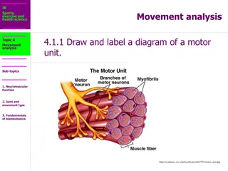
ANATOMICAL DESCRIPTION OF A NMJ Each neuromuscular junction consists of the axon terminal of a motor neuron and the motor end plate of a muscle fibre. The Motor Neuron Part: - The axon of a motor neuron enters the structure of skeletal muscle and forms many branches called axon terminals. - There is a swelling called a synaptic end bulb at the end of each axon terminal. - Each synaptic end bulb contains many synaptic vesicles each of which contains an important neurotransmitter called acetylcholine.
ANATOMICAL DESCRIPTION OF A NMJ The Muscle Fiber Part: The part of the sarcolemma of the muscle cell that is in closest proximity to the synaptic end bulb is called the motor end plate.
ANATOMICAL DESCRIPTION OF A NMJ The Synapse or Neuromuscular Junction (NMJ): The area between the axon terminal and the sarcolemma is called the 'synaptic cleft'.
NEUROMUSCULAR JUNCTION PHYSIOLOGY Release of Ach: - When a nerve pulse reaches a synaptic end bulb, it triggers release of the neurotransmitter acetylcholine (ACh) from synaptic vesicles that contain acetylcholine (ACh). - ACh then diffuses across the synaptic cleft between the motor neurone and the motor end plate.
Activation of ACh receptors: - The motor end plate contains receptors onto which the free ACh binds after diffusing across the synaptic cleft. - This binding of ACh to ACh receptors in the motor end plate causes ion channels to open & so allow the sodium (Na+) ions to flow across the membrane into the muscle cell.
NEUROMUSCULAR JUNCTION PHYSIOLOGY Generation of muscle action potential: - The flow of sodium (Na+) ions across the membrane into the muscle cell generates a muscle action potential. - This action potential then travels along the sarcolemma.
NEUROMUSCULAR JUNCTION PHYSIOLOGY Breakdown of Ach: The ACh that is released is only available to take part for a short time before it is broken down by an enzyeme called acetylcholinesterase (AChE). This breakdown of ACh occurs within the synaptic cleft.
CLASSIFICATION OF NMJ DISORDERS According to the mechanism of action or etiology: Immune-mediated disease. Toxic/metabolic. Congenital syndromes.
CLASSIFICATION Immune-mediated Myasthenia gravis, and Lambert-Eaton syndrome Toxic/metabolic Include snake venom poisoning, botulism, arthropod poisoning, organophosphates and hypermagnesemia Congenital Congenital myasthenic syndromes
CLASSIFICATION According to the location of their disruption: Presynaptic membrane of the motor neuron. The synapse. Postsynaptic membrane (the muscle fiber).
CLASSIFICATION Presynaptic Different mechanisms. Most often this causes a decrease in the release of acetylcholine. Mechanism of action can also impair the calcium channels that induce exocytosis of the vesicles. Other ion channels can also be disrupted, such as the potassium channels causing inefficient repolarization at the presynaptic membrane as in neuromyotonia. Examples: autoimmune neuromyotonia, Lambert-Eaton syndrome, congenital myasthenia gravis and botulism
CLASSIFICATION Postsynaptic The highest number of diseases affect the neuromuscular junction postsynaptically. Immune mediated Myasthenia Gravis is the most common. All the diseases that affect the postsynaptic membrane are forms of myasthenia gravis. Examples includes: Neonatal Myasthenia Gravis, Drug Induced Myasthenia Gravis and several types of Congenital myasthenia.
MYASTHENIA GRAVIS Myasthenia gravis is the most common disorder of neuromuscular transmission. The hallmark of the disorder is a fluctuating degree and variable combination of weakness in ocular, bulbar, limb, and respiratory muscles.
There are two clinical forms of myasthenia gravis: ocular and generalized. Ocular myasthenia: the weakness is limited to the eyelids and extraocular muscles. Generalized disease, the weakness commonly affects ocular muscles, but it also involves a variable combination of bulbar, limb, and respiratory muscles.
MYASTHENIA GRAVIS EPIDEMIOLOGY : Myasthenia gravis is a relatively uncommon disorder with an annual incidence of approximately 7 to 23 new cases per million. Myasthenia gravis occurs at any age, but there is a bimodal distribution to the age of onset: -Early peak in the second and third decades (female predominance) -Late peak in the sixth to eighth decade (male predominance).
PATHOPHYSIOLOGY OF MG - With every nerve impulse, the amount of ACh released by the presynaptic motor neuron normally decreases because of a temporary depletion of the presynaptic ACh stores (a phenomenon referred to as presynaptic rundown). - In MG, there is a reduction in the number of AChRs available at the muscle endplate and flattening of the postsynaptic folds. - Even if a normal amount of ACh is released, fewer endplate potentials will be produced, and they may fall below the threshold value for generation of an action potential. The end result of this process is inefficient neuromuscular transmission.
PATHOPHYSIOLOGY OF MG - Inefficient neuromuscular transmission together with the normally present presynaptic rundown phenomenon results in a progressive decrease in the amount of muscle fibers being activated by successive nerve fiber impulses. This explains the fatigability seen in MG patients - Patients become symptomatic once the number of AChRs is reduced to approximately 30% of normal.
PATHOPHYSIOLOGY OF MG - The cholinergic receptors of smooth and cardiac muscle have a different antigenicity than skeletal muscle and usually are not affected by the disease - The decrease in the number of postsynaptic AChRs is believed to be due to an autoimmune process whereby anti-AChR antibodies are produced and block the target receptors, cause an increase the turnover of the receptors, and damage the postsynaptic membrane in a complement-mediated manner.
CLINICAL FEATURES OF MG - >50% of patients present with ocular symptoms of ptosis and/or diplopia. - Of those who present with ocular manifestations, about half will develop generalized disease within two years. - 15% of patients present with bulbar symptoms. These include dysarthria, dysphagia, and fatigable chewing. -<5% present with proximal limb weakness alone.
CLINICAL FEATURES OF MG Ocular muscles: Weakness of the eyelid muscles can lead to ptosis (flactuating). The ptosis may start bilaterally and improve in one eye, resulting in unilateral ptosis or alternate. Variable severity Extraocular muscles involvement( binocular diplopia). It may be horizontal or vertical.
CLINICAL FEATURES OF MG Bulbar muscles Muscles of jaw closure (fatigable chewing). Oropharyngeal muscle weakness produces dysarthria and dysphagia. Palatal muscles weakness causing nasal speech. Nasal regurgitation, particularly of liquids, may occur due to palatal weakness
CLINICAL FEATURES OF MG Facial muscles Frequently involved and causing expressionless face. Transverse smile may be evident on examination "myasthenic sneer," where the mid-lip rises but the outer corners of the mouth fail to move. Orbicularis oculi weakness.
Neck and limb muscles : Neck extensor and flexor muscles are commonly affected. Dropped head syndrome. Proximal limb weakness (the arms > the legs). Wrist and finger extensors and foot dorsiflexors.
CLINICAL FEATURES OF MG Respiratory muscles : Respiratory muscle weakness can leads to respiratory insufficiency and pending respiratory failure "myasthenic crisis. It may occur spontaneously during an active phase of the disease or may be precipitated by a variety of factors including surgery, infections, certain medications, or tapering of immunotherapy.
DIAGNOSIS OF MG BEDSIDE TESTS: Ice pack test: It can be used in patients with ptosis. A bag (or surgical glove) is filled with ice and placed on the closed lid for two minutes. The ice is then removed and the extent of ptosis is immediately assessed. The sensitivity appears to be about 80%.
DIAGNOSIS OF MG Edrophonium(Tensilon) test: It should be used only in those patients with obvious ptosis or ophthalmoparesis, in whom improvement after infusion of the drug can easily be observed. Edrophonium chloride is an acetylcholinesterase inhibitor with rapid onset (30 to 45 seconds) and short duration of action (5 to 10 minutes). It prolongs the presence of acetylcholine in the neuromuscular junction and results in an immediate increase in muscle strength in many of the affected muscles.
DIAGNOSIS OF MG SEROLOGIC TESTING : - Acetylcholine receptor binding antibodies found in in 80-90% of those with generalized disease and in 40-55% of those with ocular myasthenia - MuSK antibodies are present in 38-50% of those with generalized myasthenia gravis who are AChR-Ab negative
DIAGNOSIS OF MG ELECTROPHYSIOLOGIC CONFIRMATION Repetitive nerve stimulation: The nerve is electrically stimulated 6 to 10 times at low rates (2 or 3 Hertz). The compound muscle action potential (CMAP) amplitude is recorded from the electrodes over the muscle after electrical stimulation of the nerve. In normal muscles, there is no change in CMAP amplitude with repetitive nerve stimulation. In myasthenia there may be a progressive decline in the CMAP amplitude with the first four to five stimuli
DIAGNOSIS OF MG An RNS study is considered positive (abnormal) if the decrement is greater than 10 % RNS studies are positive in more than 75% of patients with generalized myasthenia.
Single fiber electromyography: It is positive in greater than 90% of those with generalized myasthenia.
DIAGNOSIS OF MG CT Mediastinum: In AChR antibody positive myasthenia gravis,>75% of patients have thymic abnormalities. Thymic hyperplasia is most common 85%. Thymic tumors (primarily thymoma) in up to 15%.
DIAGNOSIS OF MG Autoimmune disorders: Autoimmune thyroid disease is common (3-8%) in patients with myasthenia. Screening for thyroid abnormalities should also be part of the initial evaluation.
TREATMENT OF MG Symptomatic treatments (anticholinesterase agents) Chronic immunotherapies (glucocorticoids/immunosuppressive drugs). Rapid immunotherapies (plasma exchange and intravenous immune globulin [IVIG]). Thymectomy.
LAMBERT EATON SYNDROME Lambert-Eaton myasthenic syndrome (LEMS) is a rare presynaptic disorder of neuromuscular transmission in which quantal release of acetylcholine (ACh) is impaired.
PATHOPHYSIOLOGY OF LEMS An autoimmune attack directed against the voltage-gated calcium channels (VGCCs) on the presynaptic motor nerve terminal results in a loss of functional VGCCs at the motor nerve terminals. The number of quanta released by a nerve impulse is diminished. Because presynaptic stores of ACh and the postsynaptic response to ACh remain intact, rapid repetitive stimulation or voluntary activation that aids in the release of quanta will raise the endplate potential above threshold and permit generation of muscle action potential.
PATHOPHYSIOLOGY OF LEMS Clinically, this phenomenon is noted by the appearance of previously absent tendon reflexes following a short period of strong muscle contraction by the patient. Parasympathetic, sympathetic, and enteric neurons are all affected
ETIOLOGY OF LEMS Autoimmune: Antibodies directed against the voltage-gated calcium channel (VGCC). These antibodies interfere with the normal calcium flux required for the release of acetylcholine. Paraneoplastic: The expression of functional VGCCs in the surface membrane of small cell lung cancer (SCLC) cells (among numerous other neural antigens) is responsible for most cases of paraneoplastic LEMS.
EPIDEMIOLOGY OF LEMS The true incidence of LEMS is unknown, but the condition is uncommon and occurs much less frequently than myasthenia gravis approximately 1/2 of LEMS cases are associated with a malignancy, mainly small cell lung cancer (SCLC) The incidence and prevalence of LEMS in patients with SCLC are estimated to be approximately 3% The other tumors associated with LEMS are lymphoproliferative disorders (Hodgkin lymphoma).
CLINICAL MANIFESTATION OF LEMS: Most patients with LEMS present with slowly progressive proximal muscle weakness, particularly involving the legs. Deep tendon reflexes are typically depressed or absent Dry mouth is the most common autonomic symptom, while erectile dysfunction is common in men Ocular symptoms, especially ptosis and diplopia, may occur with LEMS but are rarely the presenting or dominant feature of the illness.
CLINICAL MANIFESTATION OF LEMS Most patients do not have significant respiratory muscle weakness Recovery of lost deep tendon reflexes or improvement in muscle strength with vigorous, brief muscle activation is a unique aspect of LEMS
DIAGNOSIS OF LEMS : The diagnosis of LEMS is usually made on clinical grounds and confirmed by the presence of antibodies to voltage-gated calcium channel (VGCC) and by electrodiagnostic studies Antibodies against the P/Q-type VGCC are present in approximately 85-95% of patients with LEMS high frequency (10 to 50 Hz) repetitive nerve stimulation (RNS) or brief (eg, 10 seconds) maximal isometric muscle activation result in significant increment with a marked increase in the CMAP amplitude