Gel Electrophoresis in Genetic Engineering: Principles and Types

Gel electrophoresis is a crucial technique in genetic engineering used to separate macromolecules based on size and charge. Agarose and polyacrylamide gels are the two main types used, each with its advantages and disadvantages. Agarose gel electrophoresis is effective for separating large DNA molecules, while polyacrylamide gel electrophoresis is used for high-resolution techniques like sequencing. Understanding the principles and types of gel electrophoresis is essential for analyzing and manipulating DNA, RNA, and proteins in biotechnology.

Download Presentation

Please find below an Image/Link to download the presentation.
The content on the website is provided AS IS for your information and personal use only. It may not be sold, licensed, or shared on other websites without obtaining consent from the author. Download presentation by click this link. If you encounter any issues during the download, it is possible that the publisher has removed the file from their server.
E N D
Presentation Transcript
Genetic Engineering Third grade lec. 5
Principle of Gel Electrophoresis Gel electrophoresis is a macromolecules according to their size and charge. It is frequently used to analyze and manipulate samples of DNA, RNA, or proteins. Gel medium Two gel types are employed: agarose and polyacrylamide gels. Agarose gels are used as thick layers in flatbed chambers mainly for preparative purposes, whereas polyacrylamide gels are applied in thin layers in vertical or cooled flatbed systems, mainly for high resolution techniques like sequencing and genotyping ( Table 1). basic biotechnology technique that separates Table 1. Some media for electrophoresis : Medium Conditions Principal Uses Starch Cast in tubes or slabs Proteins Agarose gel Cast in tubes or slabs Very large proteins, nucleic acids, No cross-linking nucleoproteins Acrylamide gel Cast in tubes or slabs Proteins and nucleic acids Cross-linking
NOTE: Chemically, agarose is a polysaccharide, whose monomeric unit is a disaccharide of D-galactose and 3,6-anhydro-L-galactopyranose which is shown in the diagram below. Advantages of agarose gel electrophoresis: 1- Nontoxic gel medium 2- Gels are quick and easy to cast 3- Good for separating large DNA molecules 4- Can recover samples by melting the gel, digesting with enzyme agarose or treating with salts .
Disadvantages : 1. High cost of agarose 2. Fuzzy bands 3. Poor separation of low molecular weight samples . NOTE: Polyacrylamide, used mainly for SDS-PAGE, is a matrix formed from monomers of acrylamide and bis-acrylamide. In the absence of bis-acrylamide, the acrylamide would polymerize into long strands, not a porous gel. But as the diagram shows, bis-acrylamide cross-links the acrylamide chains and this is what gives rise to the formation of the porous gel matrix. The amount of crosslinking, the pore size and consequent separation properties of the gel can be controlled by varying the ratio of acrylamide to bisacrylamide.
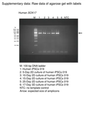
Types of gel electrophoresis: Agarose gel electrophoresis (AGE) Agarose gel electrophoresis is one of the traditional methods of separating and analyzing nucleic acid. In this method, a porous gel made from agarose acts as a separating medium. Agarose is a purified form of Agar, a gelatinous substance extracted from red algae. Agarose is in powdered form, and is insoluble in water at room temperature. Molecules with a net negative charge migrate toward the positive electrode and molecules with a net positive charge migrate toward the negative electrode. The overall charge of a molecule affects the speed at which it travels through the gel. The mobility of a molecule during gel electrophoresis also depends on its molecular size and shape. The small pores of the gel matrix act as a sieve that provides great resolving power. Small molecules move more easily through the pores than larger molecules and therefore travel relatively quickly( Figure 1). Small size and strong charge increase a molecule s migration rate through the gel. Large size and weak charge decrease the migration rate. (Note: In electrophoresis of DNA, since all the samples have the same charge, their migration rate is based only on size).The size of the fragments can be determined by running standard DNA ladder run in parallel. Migration rate of the fragments also depends on the concentration of agarose used to prepare gel (Tab 2).
Generally DNA can exist in three forms: linear form, opencircular form and supercoiled form. The linear DNA may be the product of PCR amplification or the restriction digestion product. But plasmid DNA is the one which are mostly studied. In invivo plasmids exist as highly supercoiled form to enable it to fit inside the cell. When the plasmid preparation is done plasmid DNA can exist in all the three conformations i.e. linear, opencircular and supercoiled forms (Fig. 2). Supercoiled DNA : Supercoiled DNA migrates faster than predicted in an agarose gel due to its conformation. Supercoiled DNAis the desired species when isolating plasmid DNA. Nicked, Relaxed Circular Plasmid: During replication, cellular topoisomerases nick one strand of the DNAhelix and relax the superhelical tension, thus allowing polymerases to gain access to the DNA. Nicked circle DNAis the rubber band without any twists introduced. This large floppy circle is the slowest migrating form in an agarose gel.
Linear Plasmid: Linearized DNA occurs when the DNA helix is cut in both strands at the same place. between the nicked circle and the supercoiled forms. However, it may also migrate the same distance as nicked circle. Linear DNA generally migrates Note: If you get linear DNA when you are hoping for supercoiled (e.g. after a plasmid prep) it is due to nuclease contamination or harsh treatment during purification. Circular, Single Stranded Plasmid : During alkaline lysis plasmid preps, plasmids are denatured because the hydrogen bonds are disrupted by the alkaline conditions. But the covalently-closed circular strands remain intact and topologically constrained, and when the pH is returned to neutral the hydrogen bonds reform and the supercoiled DNA is re-formed. However, if the alkaline lysis step is overly harsh (e.g. it is incubated for too long) the DNA can become permanently denatured and give you useless single stranded closed circles that migrate ahead of all of the other forms of the plasmid in a gel.
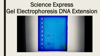
Note: Some times improper handling or storage of the isolated DNA may degrade, which can be detected as a smear when run on the agarose gel. Note: supercoiled plasmid is only one kind you want for successful cloning. Staining of the bands Ethidium bromide (EtBr) is traditionally used as a dye that binds to DNA and fluoresces under ultraviolet light. EtBr causes mutation and must be handled as hazardous waste. Due to the hazardous nature of the EtBr, recently non-toxic dyes have been introduced. While proteins separated on a polyacrylamide gel can be detected by various methods such as : Coomassie blue staining , Silver staining & Detection of radioactive proteins by autoradiography .
Recovery of DNA fragments from gels Several different procedures are used for the isolation of nucleic acids from agarose gels: electroelution, absorption to DEAE paper, absorption to glass powder or resins, digestion of agarose with enzymes.
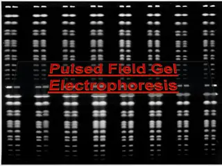
Pulsed field gel electrophoresis DNA fragments longer than about 20 kb cannot be resolved in conventional agarose gel electrophoresis because long DNA molecules align themselves as rods and migrate with a mobility that is independent of their length. In pulsed field gel electrophoresis (PFGE), the molecules are subjected to two alternating electrical fields that are applied on the gel at an angle between 110 and 180 . In PFGE, the resulting electrophoretic mobilities depend on the pulse time: DNA molecules with fragment sizes up to about 10 megabases (Mb) can be resolved. Pulse times of 1 s to 90 min are applied, depending on the length of the DNA molecules being analyzed. Large molecules are better separated with long pulse times, small molecules need short pulse times. Separations can take several days. In order to prevent chromosome-size molecules breaking by shear forces during pipetting, sample preparation including cell disruption is carried out inside little agarose blocks. These agarose blocks are inserted into preformed sample wells of the separation gel.
Polyacrylamide gel electrophoresis (SDS-PAGE) SDS-PAGE stands for Sodium Dodecyl Sulfate PolyAcrylamide Gel Electrophoresis. As with DNA agarose gel electrophoresis, in this process, proteins will be moving through polyacrylamide gels in an electrophoretic field (although nucleic acids can also be run in polyacrylamide gels instead of an agarose gel if precise separation is required). Ammonium persulfate (APS) and tetramethylethylenediamine (TEMED) are commonly used as polymerization stabilizers. SDS is used as a chemical denaturation agent, thereby unfolding the protein, and thus eliminating conformation- related mobility differences. SDS also applies a negative charge to proteins, which is crucial to further eliminate mobility differences in proteins due to charges in the amino acid side chains. In addition to SDS, protein samples are usually also heated, which helps complete unfolding, Therefore, SDS-PAGE can be used to separate proteins purely based on size (charge and conformation effects are eliminated). Visualization of proteins can be done using specific dyes such as Coomassie Brilliant Blue R-250 (commonly referred to as Coomassie blue) or silver stain.
Two-dimensional gel electrophoresis Another analytical technique in biochemistry, is 2-dimensional (2D) gel electrophoresis. The two dimensions are required for higher resolution separation of proteins: in the first dimension there is separation of nondenatured proteins (based mainly on charge, called isoelectric focusing), and in the second dimension there is a purely size-based separation in the denaturing gel (similar to SDS-PAGE). The principle is this technique: in the first dimension, the isoelectric focusing separates proteins based essentially on charge, through a gel that contains a pH gradient (which can be adjusted according to the protein groups to be studied). Proteins move along the gel to either the positive or negative end, until they reach their isoelectric point, at which point the net charge on the protein will be zero, and hence the proteins will no longer have mobility. After the first dimension, the gel (with different proteins settled at their respective isoelectric points) will be mounted onto an SDS-PAGE denaturing gel, whereby proteins from the first dimension will now be separated according to size.


")